
Study Guide for Campbell Biology
11th Edition
ISBN: 9780134443775
Author: Lisa A. Urry, Michael L. Cain, Steven A. Wasserman, Peter V. Minorsky, Jane B. Reece, Martha R. Taylor, Michael A. Pollock
Publisher: PEARSON
expand_more
expand_more
format_list_bulleted
Videos
Textbook Question
Chapter 48, Problem 3IQ
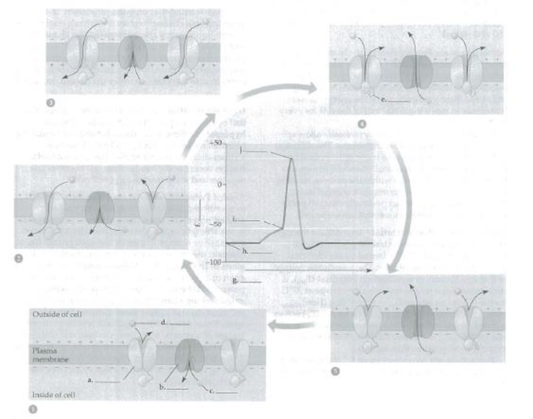
The following diagram shows the changes in voltage-gated channels during an action potential. Label the channels and inactivation loop, the ions, and the components of the graph. Name and describe the five phases of the action potential. Place numbers on the graph to show where each phase is occurring.
Expert Solution & Answer
Want to see the full answer?
Check out a sample textbook solution
Students have asked these similar questions
Name the three phases of an action potential. Describe for each the underlying molecular basis and the ion involved. Why is the term voltage-gated channel applied to Na+ channels involved in the generation of an action potential?
Graph an action potential, showing the change inelectrical potential on the y-axis and time on the x-axis.Indicate on the graph the phases when voltage-gated Na1 andK1 ion channels are opened and when they are closed
In the figure to the left, name the 4 phases of the action potential (Note: you have to write in where phase 4 occurs). Describe what happens in each phase with a focus on Na+ and K+ flow through channels and the membrane potential.
Discuss the importance of threshold. How does this relate to the concept of APs being all or none?
Chapter 48 Solutions
Study Guide for Campbell Biology
Ch. 48 - Prob. 1IQCh. 48 - a. What is the principal cation inside the cell?...Ch. 48 - The following diagram shows the changes in...Ch. 48 - Prob. 4IQCh. 48 - Prob. 5IQCh. 48 - Prob. 6IQCh. 48 - Prob. 7IQCh. 48 - Develop a flowchart or diagram or write a...Ch. 48 - Prob. 1TYKCh. 48 - Prob. 2TYK
Ch. 48 - During a neurons resting state a. there are more...Ch. 48 - Which of the following contribute(s) to the...Ch. 48 - Prob. 5TYKCh. 48 - Prob. 6TYKCh. 48 - Prob. 7TYKCh. 48 - After the rapid depolarization of an action...Ch. 48 - Nodes of Ranvier are a. gaps where Schwann cells...Ch. 48 - Prob. 10TYKCh. 48 - Signal transmission is faster in myelinated axons...Ch. 48 - Which of the following statements concerning...Ch. 48 - Prob. 13TYKCh. 48 - Prob. 14TYKCh. 48 - Prob. 15TYKCh. 48 - Prob. 16TYKCh. 48 - Prob. 17TYKCh. 48 - If the binding of a neurotransmitter to its...
Knowledge Booster


Learn more about
Need a deep-dive on the concept behind this application? Look no further. Learn more about this topic, biology and related others by exploring similar questions and additional content below.Similar questions
- Draw and label an action potential, indicating the ion movements responsible for the rising phase and the falling phase.arrow_forwardConformational changes in channel proteins brought about by voltage changes are responsible for opening and closing Na+ and K+ gates during the generation of an action potential. (True or false?)arrow_forwardDiagram an Action potential: an action potential graph showing the 4 steps of an action potential AND what is happening to the sodium voltage-gated channels and voltage-gated potassium channels at each steparrow_forward
- An action potential is considered an "all or nothing" event. What does this mean? Explain using the picture below to discuss what happens during each event. Use the letters in the diagram to match the events. +40- В -C 0- -40- E -60- -80- A 1 3 4 Time (msec) MAR 15 w MacBook Air 888 F2 F3 F4 F5 F7 FB F10 %23 24 & 3 4 6 8 %3D E T Y P 11 C V M command option Membrane potential (mV) .. ..arrow_forwardConsider the following graph of an action potential: 60- 30 membrane potential (mV) 9 -30 -60 line X -90- What is happening at point "C"? Sodium channels are opening Sodium channels are closing Potassium channels are opening Potassium channels are closing Sodium channels are closing and potassium channels are opening Sodium channels are opening and potassium channels are closing time line Oarrow_forwardConsider the following three diagrams of a nerve cell membrane. They show resting potential, depolarization, and hyperpolarization. Figure out which one is which, then draw them in the order they occur in a cell that undergoes an action potential outside + Na* inside K* Na* Nat K Nat K Na potential: -80 mV outside + Na K* Na* inside Na+ K Nat Na* K+ potential: +30 mV outside Na Na Na Na* K+ inside K* Na* Kt potential: -70 mVarrow_forward
- The voltage produced by a single nerve or muscle cell is quite small, but there are many species of fish that use multiple action potentials in series to produce significant voltages. The electric organs in these fish are composed of specialized disk-shaped cells called electrocytes. The cell at rest has the usual potential difference between the inside and the outside, but the net potential difference across the cell is zero. An electrocyte is connected to nerve fibers that initially trigger a depolarization in one side of the cell but not the other. For the very short time of this depolarization, there is a net potential difference across the cell, as shown. Stacks of these cells connected in series can produce a large total voltage. Each stack can produce a small current; for more total current, more stacks are needed, connected in parallel. In an electric eel, each electrocyte can develop a voltage of 150 mV for a short time. For a total voltage of 450 V, how many electrocytes must…arrow_forwardBased on the graph, the threshold voltage appears to be approximately. (Base your answer to this question on the graph below depicting an action potential.) +60 mV. +30 mV. 0 mV. −30 mV. −60 mV.arrow_forwardDraw the current that you would expect to flow during a voltage clamp experiment on a typical neuron. Voltages and time course are shown. Briefly explain why the currents are inward or outward. Be sure to provide scale bars. You should definitely label the Y axis so that the peak current value is obvious. Draw the Na+ current you would expect if there were physiological ionic gradients. Draw the K+ current you would expect if there are physiological ionic gradients. Draw the K+ current you would expect if the bath solution and the intracellular solution are both 125 mM.arrow_forward
- Summarize the steps in generating an action potential as a flowchart. You can make your flowchart on paper and take a picture of it, or make it electronically. Be sure you’ve included: the location in the neuron and components of the neuron involved, the types of cellular transport and ions involved, how action potentials can be stimulated and inhibited. you can get the information from this: https://youtu.be/HYLyhXRp298arrow_forward, illustrate below the phases of an action potential. Include in your figure the following:arrow_forwardGive a detailed, step-by-step description of the stages of an action potential, including a description of and explanation for the refractory periods and the rising and falling phases as well as return to rest. In your explanation, make sure to include 1) summation principles, 2) key membrane potentials (values), 3) location of voltage changes along the membrane, 4) states of the various voltage-gated channels. The more detail, the better. There are 5 main steps.arrow_forward
arrow_back_ios
SEE MORE QUESTIONS
arrow_forward_ios
Recommended textbooks for you
 Human Physiology: From Cells to Systems (MindTap ...BiologyISBN:9781285866932Author:Lauralee SherwoodPublisher:Cengage Learning
Human Physiology: From Cells to Systems (MindTap ...
Biology
ISBN:9781285866932
Author:Lauralee Sherwood
Publisher:Cengage Learning
The Cardiovascular System: An Overview; Author: Strong Medicine;https://www.youtube.com/watch?v=Wu18mpI_62s;License: Standard youtube license