
Study Guide for Campbell Biology
11th Edition
ISBN: 9780134443775
Author: Lisa A. Urry, Michael L. Cain, Steven A. Wasserman, Peter V. Minorsky, Jane B. Reece, Martha R. Taylor, Michael A. Pollock
Publisher: PEARSON
expand_more
expand_more
format_list_bulleted
Videos
Textbook Question
Chapter 44, Problem 5IQ
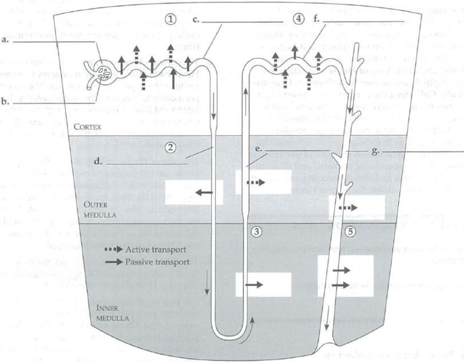
In the following diagram of a nephron and collecting duct, label the parts on the indicated lines. Review what is happening in steps 1 to 5 and label the arrows to indicate the movement of NaCl, water, nutrients, K+, HCO3−, H+, NH3, and urea into or out of the tubule.
Expert Solution & Answer
Want to see the full answer?
Check out a sample textbook solution
Students have asked these similar questions
Match the structure of the nephron to its primary function.
Renal corpuscle
Proximal tubule
Loop of Henle
Distal tubule
Collecting duct
responsible for size-selective filtration of the blood
responsible for active transport and reabsorption of ions and
nutrients as well as the reabsorption of water
may or may not reabsorb sodium ions depending on the
presence or absence of the hormone aldosterone.
establishes a strong osmotic gradient allowing the
reabsorption of water, sodium ions, and chloride ions.
regulates reabsorption of water in response to the presence of
ADH & secretes urea into the interstitial fluid to maintain the
osmotic gradient of the medulla.
The renal handling of a novel drug is being studied. When the drug is present in the blood, it is filtered into the Bowman’s capsule and secreted via transport proteins in the renal tubules, but it is NOT reabsorbed. The lines on the following graph represent filtration, secretion and excretion rates of this drug at various plasma concentrations.
For the three lines shown (labeled A-C) identify which line represents filtration, which line represents secretion, and which line represents excretion
Explain how you determined this.
Draw a schematic diagram showing the flow of water and salts in the proximal tubule of the kidney. In this diagram, put the lumen on the left side of the diagram and the extracellular fluids on the right side of the diagram. Indicate where the lumen is, where the extracellular fluid is, where the apical portion of the cell is, and show the location of the proteins that are responsible for the movement of water and salts and glucose reabsorption in this area of the kidney
Chapter 44 Solutions
Study Guide for Campbell Biology
Ch. 44 - Indicate whether the following animals are...Ch. 44 - Prob. 2IQCh. 44 - Which of the three excretory processes is least...Ch. 44 - a. What substances are removed from the blood...Ch. 44 - In the following diagram of a nephron and...Ch. 44 - a. What type of nephron enables the production of...Ch. 44 - Prob. 7IQCh. 44 - Provide the following information concerning the...Ch. 44 - Prob. 2SYKCh. 44 - Prob. 1TYK
Ch. 44 - Transport epithelia are responsible for a. pumping...Ch. 44 - Prob. 3TYKCh. 44 - Prob. 4TYKCh. 44 - Prob. 5TYKCh. 44 - Prob. 6TYKCh. 44 - Prob. 7TYKCh. 44 - Prob. 8TYKCh. 44 - Prob. 9TYKCh. 44 - The process of secretion in the formation of urine...Ch. 44 - Prob. 11TYKCh. 44 - Prob. 12TYKCh. 44 - Prob. 13TYKCh. 44 - Prob. 14TYKCh. 44 - Prob. 15TYKCh. 44 - Prob. 16TYKCh. 44 - Prob. 17TYKCh. 44 - Prob. 18TYK
Knowledge Booster


Learn more about
Need a deep-dive on the concept behind this application? Look no further. Learn more about this topic, biology and related others by exploring similar questions and additional content below.Similar questions
- As the text noted, two-thirds of the water and solutes that the body reclaims by reabsorption in nephrons occurs in the proximal tubule. Proximal tubule cells have large numbers of mitochondria and demand a great deal of oxygen. Explain why.arrow_forwardReabsorption depends on _________. a. osmosis across the nephron wall b. active transport of sodium across the nephron wall c. a steep solute concentration gradient d. all of the abovearrow_forwardDescribe (and label on a diagram) the structures of a nephron (including associated blood vessels) and give the functions of each structure.arrow_forward
- Figure 41.6 Which of the following statements about the nephron is false? The collecting duct empties into the distal convoluted tubule. The Bowman’s capsule surrounds the glomerulus. The loop of Henle is between the proximal and distal convoluted tubules. The loop of Henle empties into the distal convoluted tubule.arrow_forwardWhich of the following does not contribute to the process of filtration? (a) active transport by epithelial cells lining renal tubules (b) large surface area for filtration (c) low permeability of glomerular capillaries (d) high hydrostatic blood pressure in glomerular capillaries (e) podocytesarrow_forwardWater and small solutes enter nephrons during ________. a. filtration b. reabsorption c. secretion d. both a and barrow_forward
- Which of the following does not contribute to the high salt concentration in the interstitial fluid of the kidney medulla? (a) active transport of sodium from the upper part of the ascending limb (b) diffusion of salt from the ascending limb of the loop of Henle (c) reabsorption of salt from various regions of Bowmans capsule (d) counterflow of fluid through the two limbs of the loop of Henle (e) diffusion of urea out of the collecting ductarrow_forwardMatch each function to its site of action in the nephron. Glomerulus Proximal convoluted tubule Descending limb- of loop of Henle Answer Bank reabsorption of salt only, diluting filtrate Distal convoluted tubule Ascending limb of loop of Henle regulation of NaCl and K+ in bodily fluids by secreting K+ and reabsorbing NaCl filtration of blood p;plasma Collecting duct reabsorption of water only, increasing solute concentration of filtrate secretion of drugs and poisons into nephron lumen transport of filtrate to renal pelvisarrow_forwardSeveral pharmacological agents have been shown to lower the pressure of the filtrate inside the lumen of nephrons. What affect will this have? a hydrostatic pressure of fluid in the Bowman’s capsule increases, GFR increases b hydrostatic pressure of fluid in the Bowman’s capsule decreases, GFR decreases c hydrostatic pressure of fluid in the Bowman’s capsule increases, GFR decreases d hydrostatic pressure of fluid in the Bowman’s capsule decreases, GFR increasesarrow_forward
arrow_back_ios
arrow_forward_ios
Recommended textbooks for you
 Biology 2eBiologyISBN:9781947172517Author:Matthew Douglas, Jung Choi, Mary Ann ClarkPublisher:OpenStax
 Human Physiology: From Cells to Systems (MindTap ...BiologyISBN:9781285866932Author:Lauralee SherwoodPublisher:Cengage Learning
 Biology (MindTap Course List)BiologyISBN:9781337392938Author:Eldra Solomon, Charles Martin, Diana W. Martin, Linda R. BergPublisher:Cengage Learning
 Human Biology (MindTap Course List)BiologyISBN:9781305112100Author:Cecie Starr, Beverly McMillanPublisher:Cengage Learning
Biology 2e
Biology
ISBN:9781947172517
Author:Matthew Douglas, Jung Choi, Mary Ann Clark
Publisher:OpenStax
Human Physiology: From Cells to Systems (MindTap ...
Biology
ISBN:9781285866932
Author:Lauralee Sherwood
Publisher:Cengage Learning
Biology (MindTap Course List)
Biology
ISBN:9781337392938
Author:Eldra Solomon, Charles Martin, Diana W. Martin, Linda R. Berg
Publisher:Cengage Learning
Human Biology (MindTap Course List)
Biology
ISBN:9781305112100
Author:Cecie Starr, Beverly McMillan
Publisher:Cengage Learning
Excretory System; Author: Amoeba Sisters;https://www.youtube.com/watch?v=q5qaGHfdmYM;License: Standard youtube license