
Becker's World of the Cell (9th Edition)
9th Edition
ISBN: 9780321934925
Author: Jeff Hardin, Gregory Paul Bertoni
Publisher: PEARSON
expand_more
expand_more
format_list_bulleted
Videos
Textbook Question
Chapter 5, Problem 1Q
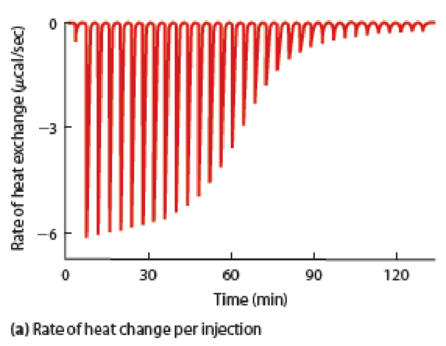
QUESTION: Why does the height of the spike get shorter after successive injections of ligand in Figure 5B-2a?
Expert Solution & Answer
Want to see the full answer?
Check out a sample textbook solution
Students have asked these similar questions
Actin alone
Actin + Protein X
--
Time
[F-actin] (µM)
The top panel (a) of this figure shows the graded potential change (far right, upper, electrical trace) that results from ligand binding to the ligand gated Na+ channel. The bottom panel of this figure (b) shows a graded potential change (far right, lower, electrical trace) that results from ligand binding to a ligand gated Cl- channel. From this trace you know (Vm = -70 mV)
1. ECl- is -70 mV
2. ECl- is more negative than -70 mV (i.e., -80 mV)
3. ECl- is more positive than -70 mV (i.e., -60 mV)
Estimate the binding affi nity of a ligand for its receptor from the followingdata:
Chapter 5 Solutions
Becker's World of the Cell (9th Edition)
Ch. 5 - How do phototrophs and chemotrophs depend...Ch. 5 - How does the spontaneity of a chemical reaction...Ch. 5 - QUESTION: Why does the height of the spike get...Ch. 5 - For a chemical reaction happening in a cell, what...Ch. 5 - Prob. 5.1PSCh. 5 - QUANTITATIVE Photosynthetic Energy Transduction....Ch. 5 - Energy Conversion. Most cellular activities...Ch. 5 - Problem Set Enthalpy, Entropy, and Free Energy....Ch. 5 - Violating the Second Law? The second law of...Ch. 5 - QUANTITATIVE The Equilibrium Constant. The...
Knowledge Booster


Learn more about
Need a deep-dive on the concept behind this application? Look no further. Learn more about this topic, biology and related others by exploring similar questions and additional content below.Similar questions
- 19:20 uil 4G A docs.google.com Observe the figure below and answer the following questions. Does "end A" represent the plus or the minus end? Explain. "A" End Your answer Identify the structure shown in this figure and indicate one of its functions.arrow_forwardIf instead of using 3.5 µM myoglobin (receptor) you used half of this (that is, 1.75 µM myoglobin), what would be that value of the Kd, that you calculated ( how would it change)? Please explain so I can solve on my own :) (How does changing concentration of the receptor in a ligand-receptor binding experiment affect the dissociation constant?)arrow_forwardPlease help me with these 2 homework problems.arrow_forward
- 2-89 The following is a block diagram for a sphingoglycolipid where the building blocks are labeled with letters and the linkages between building blocks are labeled with numbers. A 1 2 B C a. Which building blocks are fatty acid residues? b. Which building blocks are carbohydrate residues? c. Which linkages are amide linkages? d. Which linkages could involve a monosaccharide?arrow_forward= 20 nM. The rate of receptor-ligand complex formation with an A receptor-ligand complex has a dissociation constant of Ka added ligand concentration of 10 µM is 5 × 10³ s¯¹. What is the value of the reverse rate constant, k_₁ ? k_₁ = 8-1arrow_forwardAfter death, muscles become very stiff, a condition known as rigor mortis. Explain the molecular basis of rigor mortis - where in the contraction cycle is the muscle arrested? Why?arrow_forward
- problem53arrow_forwardFrom the Hill Plot below, the of the first binding event for the receptor-ligand system under study is: Q1 4 nM 10 μΜ -2 nM 2 nM Calculate the Hill Coefficient from the receptor-ligand binding data below: Q2 4 100 2 3 (077) log 10 8 6 4 2 0 -2 -4 -6 -6 -4 -2 0 2 4 log [L] (nM) 6 8 10arrow_forwardWhy is COVID-19 a man made? (Brief explanation only atleast 3-5 sentences)arrow_forward
- Crystal structures exist for three neurokinin-1 (NK1) ligand complexes with the following pdb codes (6hll, 6hlo,6hlp). State which is the highest quality crystal structure indicating the criteria you used to evaluate this.arrow_forward(ii) Considering the entropic contributions to ligand-receptor binding discuss how the strength of ligand-receptor binding for the molecule below may be affected by changing the double bond (1) to a single bond and removal of the hydroxyl group (2)arrow_forwardCrystal structures of neurokinin-1 with pdb codes 6hll, 6hlo, 6hlp. Which is the highest quality crystal structure?arrow_forward
arrow_back_ios
SEE MORE QUESTIONS
arrow_forward_ios
Recommended textbooks for you

Phlebotomy: Venipuncture Procedure; Author: Medical Lab Lady Gill;https://www.youtube.com/watch?v=LC9LABPts7M;License: Standard Youtube License