
Microbiology With Diseases By Taxonomy (6th Edition)
6th Edition
ISBN: 9780134832302
Author: Robert W. Bauman Ph.D.
Publisher: PEARSON
expand_more
expand_more
format_list_bulleted
Concept explainers
Videos
Textbook Question
Chapter 16, Problem 2VI
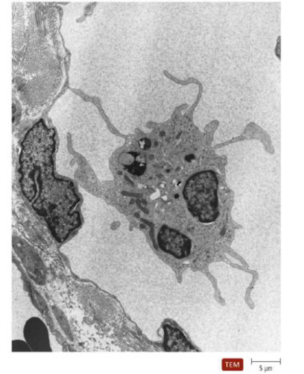
The nearby image is a transmission electron micrograph of a dendritic cell. Indicate where a scientist could find molecules of MHC I and MHC II. Label a pseudopod and a vesicle.
Expert Solution & Answer
Want to see the full answer?
Check out a sample textbook solution
Students have asked these similar questions
Can a mouse infected with Bacillus anthracis generate antibodies against the S-layer? How do you know?
I need help finding the answer in the article and explain in short answer
link to article: https://www.ncbi.nlm.nih.gov/pmc/articles/PMC106848/
Give the color reaction in Wright's stain and the function of these cells.
CELL
COLOR REACTION IN
FUNCTION
WRIGHT'S STAIN
RED BLOOD CELL
NEUTROPHIL
NUCLEUS:
CYTOPLASM/GRANULES:
EOSINOPHIL
NUCLEUS:
CYTOPLASM/GRANULES:
BASOPHIL
NUCLEUS:
CYTOPLASM/GRANULES:
LYMPHOCYTE
NUCLEUS:
CYTOPLASM/GRANULES:
MONOCYTE
NUCLEUS:
CYTOPLASM/GRANULES:
PLATELET
CYTOPLASM/GRANULES:
Write T if the statement is correct; write F if the statement is not correct.
Antigen fragments that bind to MHC class I molecules are typically in endosomes. " "
TAP1 and TAP2 are present on the plasma membrane of APCs. " "
Proteasomes process antigens within the endoplasmic reticulum. " "
Newly synthesized MHC class I molecules can be found within endosomes. " "
Newly synthesized MHC class I α chains assemble in the ER with calnexin. " "
Chapter 16 Solutions
Microbiology With Diseases By Taxonomy (6th Edition)
Ch. 16 - Prob. 1TMWCh. 16 - Prob. 2TMWCh. 16 - Prob. 3TMWCh. 16 - Prob. 4TMWCh. 16 - Prob. 5TMWCh. 16 - Prob. 1EDCSCh. 16 - Prob. 1MCCh. 16 - Prob. 2MCCh. 16 - Prob. 3MCCh. 16 - Prob. 4MC
Ch. 16 - Prob. 5MCCh. 16 - Prob. 6MCCh. 16 - Prob. 7MCCh. 16 - Which cells express MHC class I molecules in a...Ch. 16 - Prob. 9MCCh. 16 - Prob. 10MCCh. 16 - Prob. 1MTFCh. 16 - Prob. 2MTFCh. 16 - Prob. 3MTFCh. 16 - Prob. 4MTFCh. 16 - Prob. 5MTFCh. 16 - Prob. 1MCh. 16 - Prob. 2MCh. 16 - Prob. 1VICh. 16 - The nearby image is a transmission electron...Ch. 16 - Prob. 1SACh. 16 - Prob. 2SACh. 16 - Prob. 1CTCh. 16 - Contrast innate defenses with adaptive immunity.Ch. 16 - Prob. 4CTCh. 16 - Prob. 5CTCh. 16 - Prob. 6CTCh. 16 - Prob. 7CTCh. 16 - Some materials, such as metal bone pins and...Ch. 16 - Prob. 9CTCh. 16 - Prob. 10CTCh. 16 - Prob. 11CTCh. 16 - Prob. 12CTCh. 16 - Prob. 13CTCh. 16 - Prob. 14CT
Knowledge Booster


Learn more about
Need a deep-dive on the concept behind this application? Look no further. Learn more about this topic, biology and related others by exploring similar questions and additional content below.Similar questions
- The virus shown in the diagram below is only able to infect and replicate in epithelial cells. In order for the cross-presenting dendritic cell to display viral peptides, rather than self peptides on its surface MHC class I proteins, which of the following procedures could be utilized, starting with the components shown in the figure below? Mix epithelial cells with heat-killed virus, wait 24 hrs, wash away any virus particles outside the epithelial cells, then add epithelial cells to dendritic cells. Mix epithelial cells with viral peptides, wait 24 hrs, wash away any viral peptides not bound to the epithelial cells, then add epithelial cells to dendritic cells. Mix epithelial cells with live virus particles, wait 24 hrs, wash away any virus particles outside the epithelial cells, then add epithelial cells to dendritic cells. Mix dendritic cells with viral nucleic acids and epithelial cells for 24 hrs. MIx epithelial cells will viral nucleic acids, wait 24 hrs, wash away any viral…arrow_forwardA 18 month-old child is found to have measles antibodies in his blood. Suggest 3 different ways how the child could have acquired such antibodies 2. Describe what usually happens in a phagocytic vesicle after a phagocyte has engulfed a bacterium (diagrams maybe used but not required):arrow_forwardInvading microbes can be imprisoned by a ______________. Question options: blood clot cytochrome hemolysin histamine siderophore tautomer Which of the following statements is FALSE? Question options: acute inflammation develops fairly quickly chronic inflammation is harmful the permeability of small veins in an inflamed area increases the warmth in an inflamed area is a result of additional heat transported to the area by blood vasodilation increases blood flow to an area during an inflammatory response all these statements are TRUEarrow_forward
- What are MHC class I and class II receptors and how do they recognize foreignness? This is an immunology questionarrow_forwardWhich of the following sentences is CORRECT? A. P53 is an example of tumour activator gene B. leukotrienes are derived from arachidonic acid by cyclooxygenase C. effective bacteriocidal enzymes are contained in lysosome of the lymphocytes D. Gram stain is the most commonly used tissue stain for routine histological examination E. proteoglycans are located in ground substance of the ECM in connective tissues and basement membranearrow_forwardhow would you explain this article to someone to summarize the article and its sections? https://www.frontiersin.org/articles/10.3389/fimmu.2017.00292/full Major Histocompatibility Complex (MHC) Class I and MHC Class II Proteins: Conformational Plasticity in Antigen Presentationarrow_forward
- A bacterial cell has been tagged by antibodies and is destined to undergo phagocytosis. Discuss the process of phagocytosis and the steps involved. Make sure to discuss in your answer lysosomes, digestion and phagolysosomes.arrow_forwardA patient came to the health center have recurring fever, unexplained tiredness, and prolonged swelling of the lymph glands. Also, the patient’s CD4 count is less than 200 cell/mm3. Identify the organism that cause the condition also explain how you come up with the diagnosis? Provide reason why CD4 cell were affected. Give the scheme of test you would run to confirm the pathogen?arrow_forwardWhile the functions of the three types of interferons mentjoned in the text book are numerous, they all share an antiviral function. Part D- Functions and Characteristics of Cytokines Match the following functions and characteristics with the corresponding type of cytokine. Drag and drop the phrases into the bins they describe. > View Available Hint(s) Reset Help Promote secretion of adrenocorticotrophic hormone (ACTH) Stimulate blood cell production Used to treat Kaposi's sarcoma Impart viral resistance to cells that secrete this cytokine Attract free macrophages and microphages Promote activity of fibroblasts Slow growth of tumors and kills sensitive tumor cells Colony-stimulating factors Phagocyte-activating chemicals Interleukins Interferons Tumor necrosis factors Stimulate inflammatory process Stimulate the activity of NK cells Slow progress of inflammation Submit rovide Feedbackarrow_forward
- You are presented with the following clinical scenario: "A 12 month old male child presents with recurrent bacterial infections. Flow cytometry shows a complete lack of B- lymphocytes." Using your knowledge of immunology and flow cytometry, identify which dot plots represent the healthy control and patient samples in each scenario and come up with a clinical diagnosis. Note that you may need to use additional background sources to fully understand what is going on at the cellular level for each diagnosis. What is the clinical diagnosis O HIV/AIDS O COVID-19 O LAD type 1 U E CD4-FITC CD8-PE CD45-PE CD8-PE 70% 13% 52% 10% 82% 5% 27% 5% CD8-PE CD4-FITC CD20-FITC CD4-FITC 17% 38% 13% 68% Agammaglobulinaemia CD45-PE CD4-FITC CD45-PE CD45-PE 35% 3% 62% 8% 93% 22% CD15-FITC CD8-PE CD20-FITC CD15-FITC Select the flow cytometry dot plot that corresponds to the healthy control sample Select the flow cytometry dot plot that corresponds to the patient sample 62% 30% 0% 77%arrow_forwardA virulence factor that works by eliminating red blood cells and other cells and tissues, potentially including phagocytes. It creates a colorless area on a blood agar plate. DNase phagocytes O alpha-hemolysins beta-hemolysins gamma-hemolysinsarrow_forwardMHC class II molecules are found Group of answer choices on all “normal” nucleated human cells only on white blood cellsarrow_forward
arrow_back_ios
SEE MORE QUESTIONS
arrow_forward_ios
Recommended textbooks for you

 Human Physiology: From Cells to Systems (MindTap ...BiologyISBN:9781285866932Author:Lauralee SherwoodPublisher:Cengage Learning
Human Physiology: From Cells to Systems (MindTap ...
Biology
ISBN:9781285866932
Author:Lauralee Sherwood
Publisher:Cengage Learning
What is cancer? What causes cancer and how is it treated? *UPDATE*; Author: Cancer Treatment Centers of America - CTCA;https://www.youtube.com/watch?v=_N1Sk3aiSCE;License: Standard Youtube License