7" auricles BIOL2201, S24 Dissection 3- Sheep Heart Good source: https://www.youtube.com/watch?v=-ZbXiOrlFJI External Anatomy 1. Set up your dissection in a way you feel comfortable. Use the included video for a suggested method. 2. Make sure to have a camera ready to take pictures of your brain to show you completed the dissection. You will need at least 3 pictures of the eye, more are fine. You will need to identify all of the bolded structures below. 3. Identify the right and left sides of the heart. Look closely and on one side you will see a diagonal line of blood vessels that divide the heart; this line is called the interventricular sulcus. The half that includes all of the apex (pointed end) of the heart is the left side. 4. Locate the coronary arteries and veins that are on the surface of the heart. 5. Find the flaps of dark tissue on the top of the heart. These ear-like flaps are called auricles. 6. The front-most vessel is the pulmonary trunk. Place a probe or pin in the vessel to mark its place. 7. Just behind the pulmonary trunk is the aorta. Depending on how the heart was removed, you might also see a branch of the aorta called the brachiocephalic artery. Place a pin or probe in the aorta to mark its place. 8. Turn the heart so that you are looking at its dorsal side (the back). Find the large opening at the top of the heart next to the right auricle. This is the superior vena cava. Place a pin or probe in this vessel, you may also use your finger to feel the inside of the right atrium. 9. Locate another opening on the backside of the heart on the left side. This is the pulmonary vein. You can feel the inside of the right atrium by probing this opening with your finger. Place a pin or probe in the pulmonary vein opening. Internal Anatomy 1. Use a scalpel to make an incision in the heart just behind the aorta. Cut the heart in half so that the front separates from the back. This is a coronal or frontal section and it should show the inside of all chambers and the valves. 2. The chordae tendinae, also called the "heartstrings" can be found attached to the thin flaps of the tricuspid valve. They are anchored to the wall of the heart at the papillary muscle. 3. View the left atrium, left ventricle, and the mitral (bicuspid) valve. Note that the left side of the heart has a much thicker muscular wall. This can be used to determine the left and right sides of the heart. You can also identify the chordae tendinae and papillary muscle on this side of the heart. 4. Insert a probe into the aorta and observe where the probe exits the heart. You may even be able to find the small aortic semilunar valve where the aorta exits the heart. This valve doesn't have chordae tendinae and was probably broken when you probed the heart.
7" auricles BIOL2201, S24 Dissection 3- Sheep Heart Good source: https://www.youtube.com/watch?v=-ZbXiOrlFJI External Anatomy 1. Set up your dissection in a way you feel comfortable. Use the included video for a suggested method. 2. Make sure to have a camera ready to take pictures of your brain to show you completed the dissection. You will need at least 3 pictures of the eye, more are fine. You will need to identify all of the bolded structures below. 3. Identify the right and left sides of the heart. Look closely and on one side you will see a diagonal line of blood vessels that divide the heart; this line is called the interventricular sulcus. The half that includes all of the apex (pointed end) of the heart is the left side. 4. Locate the coronary arteries and veins that are on the surface of the heart. 5. Find the flaps of dark tissue on the top of the heart. These ear-like flaps are called auricles. 6. The front-most vessel is the pulmonary trunk. Place a probe or pin in the vessel to mark its place. 7. Just behind the pulmonary trunk is the aorta. Depending on how the heart was removed, you might also see a branch of the aorta called the brachiocephalic artery. Place a pin or probe in the aorta to mark its place. 8. Turn the heart so that you are looking at its dorsal side (the back). Find the large opening at the top of the heart next to the right auricle. This is the superior vena cava. Place a pin or probe in this vessel, you may also use your finger to feel the inside of the right atrium. 9. Locate another opening on the backside of the heart on the left side. This is the pulmonary vein. You can feel the inside of the right atrium by probing this opening with your finger. Place a pin or probe in the pulmonary vein opening. Internal Anatomy 1. Use a scalpel to make an incision in the heart just behind the aorta. Cut the heart in half so that the front separates from the back. This is a coronal or frontal section and it should show the inside of all chambers and the valves. 2. The chordae tendinae, also called the "heartstrings" can be found attached to the thin flaps of the tricuspid valve. They are anchored to the wall of the heart at the papillary muscle. 3. View the left atrium, left ventricle, and the mitral (bicuspid) valve. Note that the left side of the heart has a much thicker muscular wall. This can be used to determine the left and right sides of the heart. You can also identify the chordae tendinae and papillary muscle on this side of the heart. 4. Insert a probe into the aorta and observe where the probe exits the heart. You may even be able to find the small aortic semilunar valve where the aorta exits the heart. This valve doesn't have chordae tendinae and was probably broken when you probed the heart.

Surgical Tech For Surgical Tech Pos Care
5th Edition
ISBN:9781337648868
Author:Association
Publisher:Association
Chapter2: Legal Concepts, Risk Management, And Ethical Issues
Section: Chapter Questions
Problem 2.4CS

Related questions
Question
Transcribed Image Text:7"
auricles
Transcribed Image Text:BIOL2201, S24
Dissection 3- Sheep Heart
Good source: https://www.youtube.com/watch?v=-ZbXiOrlFJI
External Anatomy
1. Set up your dissection in a way you feel comfortable. Use the included video for a suggested method.
2. Make sure to have a camera ready to take pictures of your brain to show you completed the dissection. You will
need at least 3 pictures of the eye, more are fine. You will need to identify all of the bolded structures below.
3. Identify the right and left sides of the heart. Look closely and on one side you will see a diagonal line of blood
vessels that divide the heart; this line is called the interventricular sulcus. The half that includes all of the apex
(pointed end) of the heart is the left side.
4. Locate the coronary arteries and veins that are on the surface of the heart.
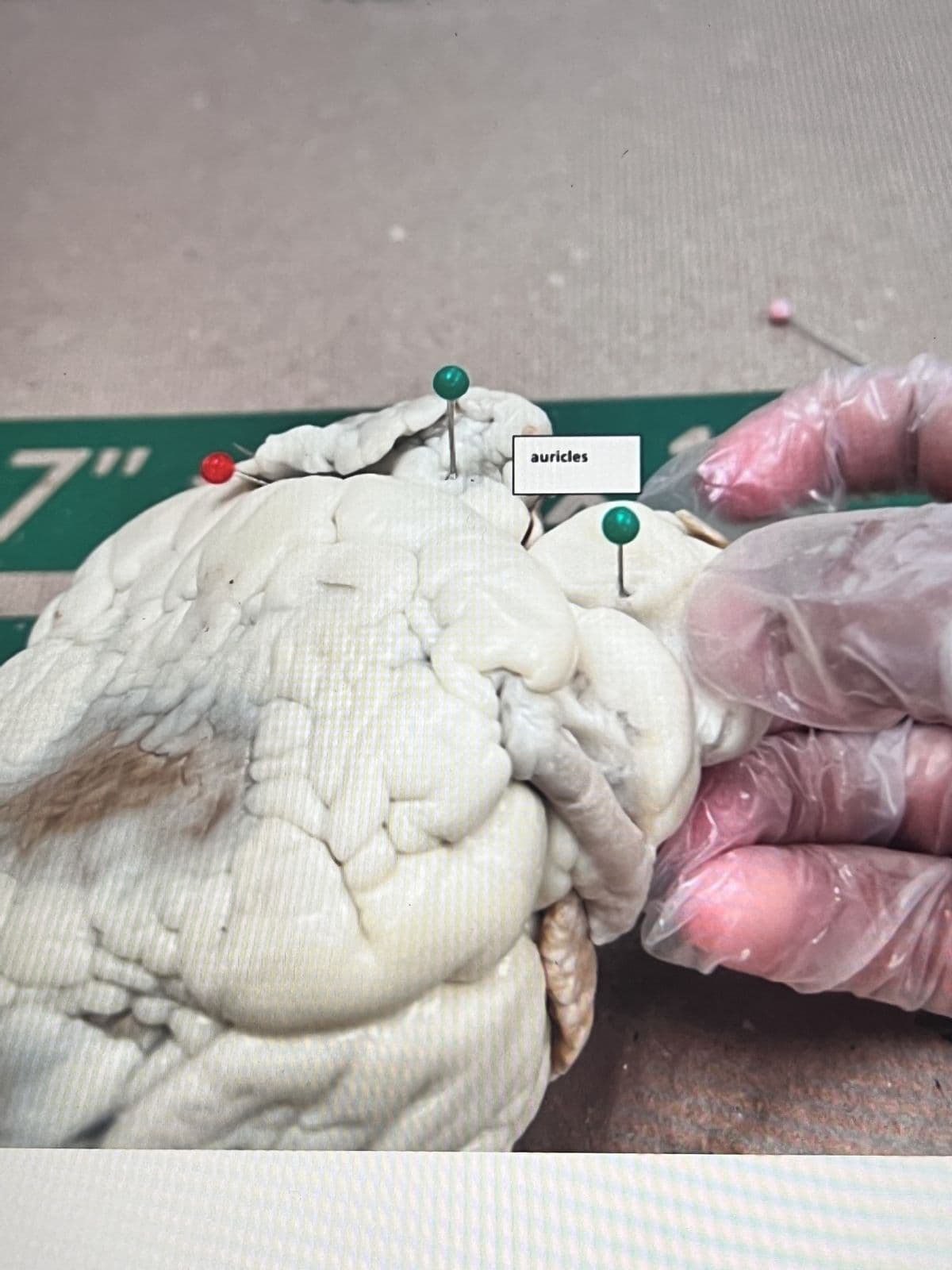
5. Find the flaps of dark tissue on the top of the heart. These ear-like flaps are called auricles.
6. The front-most vessel is the pulmonary trunk. Place a probe or pin in the vessel to mark its place.
7. Just behind the pulmonary trunk is the aorta. Depending on how the heart was removed, you might also see a
branch of the aorta called the brachiocephalic artery. Place a pin or probe in the aorta to mark its place.
8. Turn the heart so that you are looking at its dorsal side (the back). Find the large opening at the top of the heart
next to the right auricle. This is the superior vena cava. Place a pin or probe in this vessel, you may also use your
finger to feel the inside of the right atrium.
9. Locate another opening on the backside of the heart on the left side. This is the pulmonary vein. You can feel the
inside of the right atrium by probing this opening with your finger. Place a pin or probe in the pulmonary vein
opening.
Internal Anatomy
1. Use a scalpel to make an incision in the heart just behind the aorta. Cut the heart in half so that the front
separates from the back. This is a coronal or frontal section and it should show the inside of all chambers and
the valves.
2. The chordae tendinae, also called the "heartstrings" can be found attached to the thin flaps of the tricuspid
valve. They are anchored to the wall of the heart at the papillary muscle.
3. View the left atrium, left ventricle, and the mitral (bicuspid) valve. Note that the left side of the heart has a
much thicker muscular wall. This can be used to determine the left and right sides of the heart. You can also
identify the chordae tendinae and papillary muscle on this side of the heart.
4. Insert a probe into the aorta and observe where the probe exits the heart. You may even be able to find the
small aortic semilunar valve where the aorta exits the heart. This valve doesn't have chordae tendinae and was
probably broken when you probed the heart.
Expert Solution
This question has been solved!
Explore an expertly crafted, step-by-step solution for a thorough understanding of key concepts.

This is a popular solution!

Trending now
This is a popular solution!

Step by step
Solved in 2 steps

Recommended textbooks for you
Surgical Tech For Surgical Tech Pos Care
Health & Nutrition
ISBN:
9781337648868
Author:
Association
Publisher:
Cengage

Comprehensive Medical Assisting: Administrative a…
Nursing
ISBN:
9781305964792
Author:
Wilburta Q. Lindh, Carol D. Tamparo, Barbara M. Dahl, Julie Morris, Cindy Correa
Publisher:
Cengage Learning
Surgical Tech For Surgical Tech Pos Care
Health & Nutrition
ISBN:
9781337648868
Author:
Association
Publisher:
Cengage
Comprehensive Medical Assisting: Administrative a…
Nursing
ISBN:
9781305964792
Author:
Wilburta Q. Lindh, Carol D. Tamparo, Barbara M. Dahl, Julie Morris, Cindy Correa
Publisher:
Cengage Learning