Biochemistry
9th Edition
ISBN: 9781319114671
Author: Lubert Stryer, Jeremy M. Berg, John L. Tymoczko, Gregory J. Gatto Jr.
Publisher: W. H. Freeman
expand_more
expand_more
format_list_bulleted
Concept explainers
Question
Assume a 5250 base pair, closed circular plasmid with 10 negative
supercoils.
(a) Calculate the values of twist (T), writhe (W), and linking number (L) for this
plasmid (use 10.5 base pairs/turn for B-DNA)
(b) Your starting plasmid is now A-DNA instead of B-DNA. What is the linking
number required to have 10 negative supercoils in this plasmid?
(R = 8.3145 J−1 K mol−1; 0 ◦C = 273.15 K; Faraday Constant , F =96485 J V−1 mol−1.)
Expert Solution
This question has been solved!
Explore an expertly crafted, step-by-step solution for a thorough understanding of key concepts.
This is a popular solution
Trending nowThis is a popular solution!
Step by stepSolved in 4 steps

Knowledge Booster
Learn more about
Need a deep-dive on the concept behind this application? Look no further. Learn more about this topic, biochemistry and related others by exploring similar questions and additional content below.Similar questions
- A number of yeast-derived elements were added to thecircular bacterial plasmid pBR322. Yeast that requireuracil for growth (Ura− cells) were transformed withthese modified plasmids and Ura+ colonies were selected by growth in media lacking uracil. For plasmidscontaining each of the elements listed in parts (a) to(c), indicate whether you expect the plasmid to integrate into a chromosome by recombination, or insteadwhether it is maintained separately as a plasmid. If theplasmid is maintained autonomously, is it stably inherited by all of the daughter cells of subsequent generations when you no longer select for Ura+ cells (that is,when you grow the yeast in media containing uracil)?a. URA+ geneb. URA+ gene, ARS c. URA+ gene, ARS, CEN (centromere)d. What would need to be added in order for these sequences to be maintained stably in yeast cells as alinear artificial chromosome?arrow_forwardYou add 3.0 µg of plasmid DNA to a QIAprep spin miniprep purification column. You perform the binding in a binding buffer at pH 6.8. What portion (%) of the 3.0 µg of plasmid DNA would you expect to bind to the column in these conditions? What portion (%) of the bound plasmid DNA would you expect to elute using elution buffer at pH 8.5? Make the following assumptions:• Solid phase silanol groups can have a very wide range of pKas. For the purpose of this problem, assume the silica gel functional group (silanol) in the DNA purification column matrix has a pKa of 7.5.• If a DNA molecule and column matrix functional groups have the same charge state, then that DNA molecule will be repelled and will not bind to the column. If they do not have the same charge, binding occurs.arrow_forwardYou want to digest 1 µg of plasmid DNA in a final volume of 50 μL. Your solution containing plasmid has a concentration of 25 ng/μL. How many μL of your plasmid solution do you need to add to your reaction tube to digest your desired mass of plasmid?arrow_forward
- How many fragments and how big each fragment should be after digesting the plasmid with BamHI? How many fragments and how big each fragment should be after digesting the plasmid with PvuI? How many fragments and how big each fragment should be after digesting with both enzymes? Show your calculation/explanation.arrow_forwardCorrect order ib which the following enzynes would operate to fix a damaged nucleotide in a human gene. a) nuclease, DNA polymerase, RNA primase b) helicase, DNA polymerase, DNA ligase c) DNA ligase, nuclease, helicase d) nuclease, DNA polymerase, DNA ligasearrow_forwardUsing the figure below, what is molecule "A" (type a 1, 2 or 3 in the blank) nuclease ligase DNA polymerase What is the function of molecule "A"? to separate the double helix into two to piece together the Okazaki segments to copy the new DNA strand to the old strand by complementary base pairing Using the figure below, what is molecule "G" (type a 1, 2 or 3 in the blank) nuclease ligase DNA polymerase What is the function of molecule "G"? to separate the double helix into two to piecearrow_forward
- Below is a depiction of a replication bubble. 5' AGCTCCGATCGCGTAACTTT 3' TCGAGGCTAGCGCATTGAAA CTAAAGCTTCGGGCATTATCG 3' GATTTCGAAGCCCGTAATAGC TATCGACS Consider the following primer which binds to the DNA replication bubble on the diagram above: 5'-GCUAUCG-3' Identify the DNA sequence to which this primer would bind and the orientation. If the replication fork moves to the right, will the primer be used to create the leading strand of replication or the lagging strand? Explain your answer b. If the replication fork moves to the left, will the primer be used to create the leading strand of replication or a. the lagging strand? Explain your answer. What would the next five nucleotides added to the primer by DNA polymerase? С.arrow_forwardand set up a series of four dideoxy reactions (the dideoxy nucleotides are labeled with radioactive P32 ). You then separate the products of the reactions by gel electrophoresis and obtain the following banding pattern: a) Write out the base sequence of the newly synthesized strand from reading the gel (include the 5’ and 3’; end) b) Write out the base sequence of the original fragment you were given (include the 5’ and 3’ end)arrow_forwardCan you please help with 1f please picture with 1 graph is for question 1a) picture with 4 graphs is for question 1b) 1a) E. coli DNA and binturong DNA are both 50% G-C. If you randomly shear E. coli DNA into 1000 bp fragments and put it through density gradient equilibrium centrifugation, you will find that all the DNA bands at the same place in the gradient, and if you graph the distribution of DNA fragments in the gradient you will get a single peak (see below). If you perform the same experiment with binturong DNA, you will find that a small fraction of the DNA fragments band separately in the gradient (at a different density) and give rise to a small "satellite" peak on a graph of the distribution of DNA fragments in the gradient (see below). Why do these two DNA samples give different results, when they're both 50% G-C? 1b) If you denatured the random 1000 bp fragments of binturong DNA that you produced in question 1a by heating them to 95ºC, and then cooled them down to 60ºC and…arrow_forward
- A 2.0kb bacterial plasmid ‘BS1030’ is digested with the restriction endonuclease Sau3A; the plasmid map is depicted in the diagram below and the Sau3A (S) restriction sites are indicated. Which of the following DNA fragments do you expect to see on an agarose gel when you run Sau3A-digested plasmid ‘BS1030’ DNA? a. 250 bp, 450 bp, 550 bp, 1.1 kb, 1.5 kb and 2.0 kb b. 2.0kb c. 250 bp, 400 bp, 450 bp, 500 bp and 550 bp d. 100 bp, 200 bp, 250 bp, 400 bp, 500 bp and 550 bparrow_forwardThe transformation results below were obtained with 10 ul of intact plasmid DNA at nine concentrations. The following numbers of colonies are obtained when 100 ul of transformed cells are plated on selective medium: Fill in the following table: Concentration # colonies DNA mass of Fraction of Mass Transformation PGREEN (Concentration x volume OR X spread = x 10ul plasmid solution) PGREEN in cell Cell efficiency Y÷ A suspension suspension spread = 100 ul - total vol cell susp. (Colonies - Mass spread) C x Z = A See (510 ul) HINT: this calculation is constant Given= X Given=Y С. Z. 0.00001 ug/ul | 4 0.00005 ug/ul 12 0.0001 ug/ul 0.0005 ug/ul 32 125 0.001 ug/ul 442 0.005 µg/ul 0.01 ug/ul 0.05 ug/ul 0.1 ug/ul 542 507 475 516 0.5 ug/ul 505arrow_forwardAssume that a plasmid is 4700 base pairs in length and has restriction sites for a given restriction enzyme at the following locations: 800, 1400, 2900, and 3600. List the fragments by size that are ! expected when the plasmid is fully digested the restriction enzyme.arrow_forward
arrow_back_ios
SEE MORE QUESTIONS
arrow_forward_ios
Recommended textbooks for you
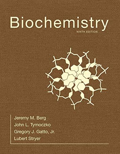
 BiochemistryBiochemistryISBN:9781319114671Author:Lubert Stryer, Jeremy M. Berg, John L. Tymoczko, Gregory J. Gatto Jr.Publisher:W. H. Freeman
 Lehninger Principles of BiochemistryBiochemistryISBN:9781464126116Author:David L. Nelson, Michael M. CoxPublisher:W. H. Freeman
 Fundamentals of Biochemistry: Life at the Molecul...BiochemistryISBN:9781118918401Author:Donald Voet, Judith G. Voet, Charlotte W. PrattPublisher:WILEY
 BiochemistryBiochemistryISBN:9781305961135Author:Mary K. Campbell, Shawn O. Farrell, Owen M. McDougalPublisher:Cengage Learning
 BiochemistryBiochemistryISBN:9781305577206Author:Reginald H. Garrett, Charles M. GrishamPublisher:Cengage Learning
 Fundamentals of General, Organic, and Biological ...BiochemistryISBN:9780134015187Author:John E. McMurry, David S. Ballantine, Carl A. Hoeger, Virginia E. PetersonPublisher:PEARSON

Biochemistry
Biochemistry
ISBN:9781319114671
Author:Lubert Stryer, Jeremy M. Berg, John L. Tymoczko, Gregory J. Gatto Jr.
Publisher:W. H. Freeman

Lehninger Principles of Biochemistry
Biochemistry
ISBN:9781464126116
Author:David L. Nelson, Michael M. Cox
Publisher:W. H. Freeman

Fundamentals of Biochemistry: Life at the Molecul...
Biochemistry
ISBN:9781118918401
Author:Donald Voet, Judith G. Voet, Charlotte W. Pratt
Publisher:WILEY

Biochemistry
Biochemistry
ISBN:9781305961135
Author:Mary K. Campbell, Shawn O. Farrell, Owen M. McDougal
Publisher:Cengage Learning

Biochemistry
Biochemistry
ISBN:9781305577206
Author:Reginald H. Garrett, Charles M. Grisham
Publisher:Cengage Learning

Fundamentals of General, Organic, and Biological ...
Biochemistry
ISBN:9780134015187
Author:John E. McMurry, David S. Ballantine, Carl A. Hoeger, Virginia E. Peterson
Publisher:PEARSON