Campbell Essential Biology (7th Edition)
7th Edition
ISBN: 9780134765037
Author: Eric J. Simon, Jean L. Dickey, Jane B. Reece
Publisher: PEARSON
expand_more
expand_more
format_list_bulleted
Concept explainers
Videos
Textbook Question
Chapter 8, Problem 16PS
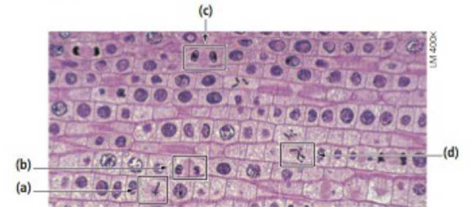
You prepare a slide with a thin slice of an onion root tip. You see the following view in a light microscope. Identify the stage of mitosis for each of the outlined cells, a–d
Expert Solution & Answer
Want to see the full answer?
Check out a sample textbook solution
Students have asked these similar questions
You prepare a slide with a thin slice of an onion root tip. You see the following view in a light microscope. Identify the stage of mitosis for each of the outlined cells, a–d
List in each blank the description of events associated with each stage of mitosis in addition to the preparatory stage Interphase. Some events and structures occur only in plant cells and some in animal cells. Mark these events in the list with an asterisk.
Draw or explain in a simple manner what needs to be done in each step of modeling mitosis.
Steps/ Procedure:
1. Tie the ends of a 10-ft (3-m) piece of string together and form a circle on the table. This represents the cell membrane of a parent cell.
2. Make a smaller circle from a 5-ft (1.5-m) piece of string to create a nuclear membrane, and place it inside the cell membrane.
3. Place 1 sock of each pair into its mate and jumble the socks inside the nuclear membrane.
4. During interphase, the cell prepares for nuclear division and DNA replicates. To simulate replication, remove a mate from each pair of socks. Match sister chromatids and connect them at their centromere by tying the matching pairs together at their centers with a small piece of string. Place them back into the nuclear membrane, jumbling them up.
5. Mitotic division now begins with prophase. During this phase, DNA condenses into chromosomes made up of 2 identical sister chromatids, and the nuclear membrane breaks…
Chapter 8 Solutions
Campbell Essential Biology (7th Edition)
Ch. 8 - Which of the following is not a function of...Ch. 8 - In what sense are the daughter cells produced by...Ch. 8 - Why is it hard to observe individual chromosomes...Ch. 8 - A biochemist measures the amount of DNA in cells...Ch. 8 - What phases of mitosis are opposite in terms of...Ch. 8 - Complete the following table to compare mitosis...Ch. 8 - If an intestinal cell in a dog contains 78...Ch. 8 - A micrograph of a dividing cell from a mouse shows...Ch. 8 - Prob. 9SQCh. 8 - Prob. 10SQ
Ch. 8 - Although nondisjunction is a random event, there...Ch. 8 - Prob. 12IMTCh. 8 - Prob. 13IMTCh. 8 - For each statement, identify which major theme is...Ch. 8 - A mule is the offspring of a horse and a donkey. A...Ch. 8 - You prepare a slide with a thin slice of an onion...Ch. 8 - Interpreting Data The graph shows the incidence of...Ch. 8 - If an endangered species can reproduce by...Ch. 8 - Every year, about a million Americans are...Ch. 8 - The practice of buying and selling gametes,...Ch. 8 - Prob. 21BS
Additional Science Textbook Solutions
Find more solutions based on key concepts
Sea turtles have disappeared from many regions, and one way of trying to save them is to reintroduce them to ar...
Marine Biology (Botany, Zoology, Ecology and Evolution)
What are the cervical and lumbar enlargements?
Principles of Anatomy and Physiology
Visit this site (http://openstaxcollege.org/l/heartvalve) to observe an echocardiogram of actual heart valves o...
Anatomy & Physiology
What are the cervical and lumbar enlargements?
Principles of Anatomy and Physiology
2. A gene is a segment of DNA that has the information to produce a functional product. The functional product ...
Genetics: Analysis and Principles
WHAT IF? As a cell begins the process of dividing, its chromosomes become shorter, thicker, and individually vi...
Campbell Biology in Focus (2nd Edition)
Knowledge Booster


Learn more about
Need a deep-dive on the concept behind this application? Look no further. Learn more about this topic, biology and related others by exploring similar questions and additional content below.Similar questions
- Using the data below for Allium cepa, express the total number of cells in mitosis as a percentage of the total number of cells in the field of view for Field 1. This is the mitotic index. Give your answer as a whole number (i.e. no decimals) and without any units. Total number of cells in Number of cells in: A.cepa root tip field of view M A Field 1 181 16 9 4 147 112 Field 2 12 7 6 Field 3 11 9arrow_forwardDraw various stages of mitosis of a cell with 4 chromosomes (2n = 4) on a piece of paper.arrow_forwardLook at the following figure and, without using your textbook or other resources, write the appropriate stage of the cell cycle after the letters A–D. Then write the appropriate stages of mitosis after the numbers 1–4. Describe what is happening at each stage.arrow_forward
- Look at the following figure and, without using your textbook or other resources, write the appropriate stage of the cell cycle after the letters A–D. Then write the appropriate stages of mitosis after the numbers 1–4.arrow_forwardPart A. Figure 1. Cells in various stages of mitotic division of an onion root tip.arrow_forwardThe following diagram shows a simulated microscopic view of a tissue sample taken from a patient where the cells have been squashed onto a slide and stained to visualize the cell's DNA. In the diagram, click or tap on the center of all cells that appear to be involved in any of the four stages of mitosis. Make sure to mark as close to the center of the cell as possible. In the diagram from Question 5, count the total number of cells visible and write the total number of cells in the space below: In the diagram from Question 5, count the number of cells that you had marked as currently undergoing any of the four phases of mitosis. How many cells in total are currently undergoing mitosis? Write this number in the space below: The mitotic index is a calculated value that represents the percentage of cells in a sample that are actively dividing. It involves counting the total number of cells present including those actively dividing or those in interphase, the total number of cells that…arrow_forward
- The phases of mitosis are shown in Figure 6.4. Mitosis is the type of nuclear division that occurs when an animal or plant grows larger and when injury heals. Two daughter cells result because there is only one round of division, and it keeps the chromosome number constant (same as the parent cell). The prophase cell in Figure 6.4 has the same number of chromosomes as the telophase nuclei in Figure 6.4. Explain the different appearance of the chromosomes.arrow_forwardThe image below is of a stained onion root tip. Determine the phase of mitosis that each cell below is in, and record that below. Look closely - cells that are clearly stained are numbered in the image below. Use those numbers when assigning the phase of mitosis for each cell. Using the same onion root tip image and data from the question above, which phase are the majority of cells in? Make a conclusion about what this means about the length of each phase of the cell cycle.arrow_forward1) What phase of mitosis is this? Answer: 2) What is the indicated structure 1? select one. a. chromosome b. chromosome with two sister chromatids c. centrioles/centrosome d. spindle fibres e. metaphase plate 3)If the diploid (2n) number of this cell is 8 how many chromosomes are present? 4) If the diploid (2n) number of this cell is 8 how many sister chromatids are present? 5) If the diploid (2n) number of this cell is 8 how many homologous pairs are present?arrow_forward
- MIosis Method: Observations of onion root tip squash. Scan the microscope under the 10x objective. Look for the region that has large nuclei relative to the size of the cell; among these cells will be found cells displaying stages of mitosis. Switch to the 40X objective to make closer observations. Since prophase and prometaphase are difficult to distinguish, classify all these cells as prophase. Record your observations in the table provided. Examples are shown in the figure below. Interphase Early Prophase Mid Prophase 10 Metaphase Anaphase Telophase Exercises: 1. Find and draw a cell showing each stage of mitosis. Interphase Prophase Metaphase Anaphase Telophase , 2. What is a distinguishing visible feature of each stage of mitosis? Interphase: Prophase: Metaphase: Anaphase: Telophase: Many of the cells of the root meristem are not undergoing mitosis; rather they Based upon the interpretations 3. are in a stage called 21arrow_forwardDetermine whether the following figures portray either Mitosis or Meiosis and identify its specific stage of cell division. (e.g. Mitosis- Prophase)arrow_forwardFrom the given picture locate and label the different stages of mitosis (prophase, metaphase, anaphase and telophase)arrow_forward
arrow_back_ios
SEE MORE QUESTIONS
arrow_forward_ios
Recommended textbooks for you
 Human Heredity: Principles and Issues (MindTap Co...BiologyISBN:9781305251052Author:Michael CummingsPublisher:Cengage Learning
Human Heredity: Principles and Issues (MindTap Co...
Biology
ISBN:9781305251052
Author:Michael Cummings
Publisher:Cengage Learning
The Cell Cycle and its Regulation; Author: Professor Dave Explains;https://www.youtube.com/watch?v=eqJqhA8HSJ0;License: Standard YouTube License, CC-BY
Cell Division - Mitosis and Meiosis - GCSE Biology (9-1); Author: Mr Exham Biology;https://www.youtube.com/watch?v=w7vp_uRA8kw;License: Standard YouTube License, CC-BY