Foundations in Microbiology
10th Edition
ISBN: 9781259705212
Author: Kathleen Park Talaro, Barry Chess Instructor
Publisher: McGraw-Hill Education
expand_more
expand_more
format_list_bulleted
Concept explainers
Videos
Textbook Question
Chapter 19.L2, Problem 2VC
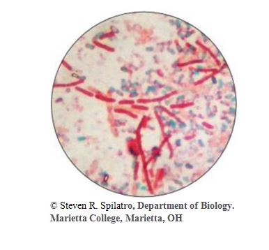
2. From chapter 3, figure 3.8. What type of bacterial infection could be diagnosed using the stain shown in the Figure? What does this result indicate?
Expert Solution & Answer
Want to see the full answer?
Check out a sample textbook solution
Students have asked these similar questions
Part A.
1.Why do microbiologists use cultured plates with 30 to 300 colonies used for calculations?
2. Why is ground beef a better bacterial growth medium than a steak or roast.
3. Why does repeated freezing and thawing increase bacterial growth if meat is then left at room temperature?
1. You accidentally used safranin as the primary stain and malachite green as the counter stain during a spore staining procedure. Explain how your microscopic observation would differ from those observed when slides of spore-containing bacteria were prepared correctly.
2.One of your classmates performed a gram stain on Pseudomonas aeruginosa and found variable gram staining, with only some of the cells gram positive. What should your classmate have found? What errors could have contributed to the variable result?
How does the streak plate technique help in isolating individual colonies of bacteria? Why streak multiple times on one plate?
3. While observing a plate inoculated with bacteria you observe a white-tan fuzzy growth. What can you interpret from this observation? Explain how this outcome could’ve occurred.
4. What are 3 possible errors that could take place as you culture your samples which could lead to contamination? State 3 and explain why.
Microbiology class
Chapter 19 Solutions
Foundations in Microbiology
Ch. 19.1 - 1. Describe how cellular characteristics are used...Ch. 19.1 - 1. Explain why Bacillus, Clostridium, and...Ch. 19.2 - 2. Recall the general characteristics of the genus...Ch. 19.2 - 3. Distinguish between cutaneous and pulmonary...Ch. 19.2 - 4. State the general characteristics of the genus...Ch. 19.2 - 5. Recall the organisms responsible for...Ch. 19.2 - Prob. 6ELOCh. 19.2 - Prob. 7ELOCh. 19.2 - Prob. 8ELOCh. 19.2 - 9. Compare food intoxication caused by Bacillus...
Ch. 19.2 - Prob. 2CYPCh. 19.2 - Prob. 3CYPCh. 19.2 - 4. What are the common elements of puncture...Ch. 19.2 - 5. What is the relationship between the normal...Ch. 19.2 - Prob. 6CYPCh. 19.2 - Prob. 7CYPCh. 19.2 - 8. ln what way is the ingested agent responsible...Ch. 19.3 - 10. Relate the severity of listeriosis with the...Ch. 19.3 - 11. Explain why people in certain occupations are...Ch. 19.3 - 9. Compare the effects of listeriosis in healthy...Ch. 19.3 - 10. Why do erysipeloids commonly appear on the...Ch. 19.4 - Prob. 12ELOCh. 19.4 - Prob. 13ELOCh. 19.4 - Prob. 11CYPCh. 19.4 - Prob. 12CYPCh. 19.5 - Prob. 14ELOCh. 19.5 - Prob. 15ELOCh. 19.5 - Prob. 16ELOCh. 19.5 - Prob. 17ELOCh. 19.5 - 18. Explain the significance of nontuberculous...Ch. 19.5 - Prob. 13CYPCh. 19.5 - 14. Compile a list of the advantages,...Ch. 19.5 - 15. Explain how and why antibacterial treatment...Ch. 19.5 - 16. List several differences between lepromatous...Ch. 19.5 - Prob. 17CYPCh. 19.5 - 18. List the diseases and at-risk populations...Ch. 19.6 - Prob. 19ELOCh. 19.6 - 20. Describe the types of infections attributable...Ch. 19.6 - 19. Compare the types of infections caused by the...Ch. 19.L1 - 1. What is/are the usual habitat(s) of...Ch. 19.L1 - Prob. 2MCQCh. 19.L1 - Prob. 3MCQCh. 19.L1 - 4. Clostridium perfringens causes a. myonecrosis...Ch. 19.L1 - Prob. 5MCQCh. 19.L1 - Prob. 6MCQCh. 19.L1 - Prob. 7MCQCh. 19.L1 - Prob. 8MCQCh. 19.L1 - Prob. 9MCQCh. 19.L1 - 10. Soil mycobacteria can be the cause of a....Ch. 19.L1 - Prob. 11MCQCh. 19.L1 - Prob. 12MCQCh. 19.L1 - Prob. 13MCQCh. 19.L1 - Prob. 14MCQCh. 19.L1 - Prob. 15MCQCh. 19.L1 - 16. Matching. Match the disease with the principal...Ch. 19.L1 - Prob. 1CSRCh. 19.L1 - 2. During this outbreak, some people sickened with...Ch. 19.L1 - 3. No listeria monocytogenes was discovered in the...Ch. 19.L1 - Prob. 1WCCh. 19.L1 - Prob. 2WCCh. 19.L1 - Prob. 3WCCh. 19.L1 - Prob. 4WCCh. 19.L1 - Prob. 5WCCh. 19.L1 - 6. a. Why is listeriosis a serious problem even...Ch. 19.L1 - Prob. 7WCCh. 19.L1 - Prob. 8WCCh. 19.L1 - 9. a. Outline the unique characteristics of...Ch. 19.L1 - Prob. 10WCCh. 19.L1 - 11. a. What is the importance of NTM? b. Describe...Ch. 19.L1 - Prob. 12WCCh. 19.L2 - Prob. 1CTCh. 19.L2 - 2. a. Why is it unlikely that diseases such as...Ch. 19.L2 - Prob. 3CTCh. 19.L2 - Prob. 4CTCh. 19.L2 - Prob. 5CTCh. 19.L2 - 6. Adequate cooking is the usual way to prevent...Ch. 19.L2 - 7. a. Why do patients who survive tetanus and...Ch. 19.L2 - Prob. 8CTCh. 19.L2 - 9. How can one tell that acne involves an...Ch. 19.L2 - Prob. 10CTCh. 19.L2 - Prob. 11CTCh. 19.L2 - Prob. 12CTCh. 19.L2 - 13. Which diseases discussed in this chapter have...Ch. 19.L2 - 14. Eighty-six people at a St. Patrick's Day...Ch. 19.L2 - 15. An outbreak of gastrointestinal illness was...Ch. 19.L2 - Prob. 1VCCh. 19.L2 - 2. From chapter 3, figure 3.8. What type of...
Knowledge Booster


Learn more about
Need a deep-dive on the concept behind this application? Look no further. Learn more about this topic, biology and related others by exploring similar questions and additional content below.Similar questions
- What is the purpose of fixing a smear? Mark all that apply: 1. To attach the bacteria to the slide 2. To cause the cells to shrink and become distorted 3. To kill the bacteria so they aren't harmed by the staining method 4. To break down the cell wall in order to make the cells accept stain 5. To kill the bacteria to make the slide safer to handlearrow_forward1 Figure 3. Two different types of bacterial colonies on an agar plate. Using microbiology terms, describe fully the colonial morphology of the two colonies shown above. A full description will include texture, transparency, color, and form (size, overall shape, margin, and elevation). Colony 1 Colony 2 2.arrow_forwardThis is how a hanging-drop slide is prepared. Figure from Macedo, Wikimedia Commons, 2016. 2 3 4 slide cover slip vaseline concavity drop of microbiological culture CA inoculation loop 11. Which slide gives you more information, the hanging drop or the stained slide? Why do you say so? 12. Why might it be valuable to know whether a bacterium is motile?arrow_forward
- a. After conducting the Gram staining procedures and you observed that all cells are stained with too much purple though the bacteria you fixed is Gram negative, what could be the mistake you have done? b. After conducting the Gram staining procedures and you observed that all cells are stained with too much pink though the bacteria you fixed is Gram positive, what could be the mistake you have done?arrow_forward8. Pigment or color (e.g.: opaque, translucid, red, yellow, rose, violet, etc..) is also another characteristic that can be used to identify your microorganism in a slant Thus, write down the name of three microorganisms (using scientific notation: Escherichia coli) that present a pigmented pattern of growth in a slant (write down the color of each pigment) and the name of the disease that they may cause. F10 F12 F6 %24 6. R. Home Enter K F G A Shift C Page Up Page Down Alt Ctrl Alt Σarrow_forward1. The colonies on a negative MSA plate would appear _____________. 2. "E. Coli and S. epidermidis were chosen to represent Gram-negative and Gram-positive bacteria, respectively. For a given antibiotic, is there a difference in susceptibility between the Gram-positive and Gram-negative bacteria?" "No, they are usually similar" "yes, drugs that target the cell membrane are more effective on gram negatives" "yes, drugs that target the cell wall are more effective on gram negatives" "yes, drugs that target the cell wall are more effective on gram positives" how can I tell the differencearrow_forward
- 3. Draw the specimen observed under endospore staining 4. Draw the specimen observed under positive simple stainingarrow_forward1. What is the most common sterilization technique used in laboratories? 2. List at least 5 procedures of the aseptic technique and describe its uses. 3. Why is Gram stain one of the most important and widely used stains in bacteriology? 4. Explain the Gram staining technique in chronological order. Indicate the reagent used and the time of usage on each reagent.arrow_forward3. A bacterial culture was diluted and results from duplicate plates were obtained as indicated below. What was the number of colony forming units/mL of the original culture? Dilution used for plating 10-2 10-3 104 10-5 10⁰ 10-7 10-8 4. 5. Amount plated 0.1 mL 0.1 mL 0.1 mL 0.1 mL 0.1 mL 0.1 mL 0.1 mL Colony counts after incubation (Results from duplicate plates) Too many to count Too many to count 341 413 99 175 27 : 29 7:2 0:0 Ten grams of hamburger were added to 90 mL of sterile buffer. This was mixed well in a blender. One-tenth of a mL of this slurry was added to 9.9 mL of sterile buffer. After thorough mixing, this suspension was further diluted by successive 1/100 and 1/10 dilutions. One-tenth of a mL of this final solution was plated onto Plate Count agar. After incubation 145 colonies were present. How many colony-forming units were present in the total 10 gram sample of hamburger? Devise a serial dilution scheme to prepare a 10-5 dilution on a plate using the least number of…arrow_forward
- What is the purpose of fixing a smear? Mark all that apply: To kill the bacteria so they aren't harmed by the staining method O To kill the bacteria to make the slide safer to handle O To attach the bacteria to the slide To break down the cell wall in order to make the cells accept stain To cause the cells to shrink and become distorted Microsoft Bing 11:30 AM do 87 F Sunny 9/14/2021arrow_forwardAnswer this question xi) What is the most important step in the Gram staining procedure? Why?arrow_forwardStep #1 Culture and perform a Gram-stain on the Super Bug You culture the Super Bug by swabbing the patient's wound, applying the sample to the surface of an agar plate (petri dish), and incubating the plate for one day. Next you perform a Gram stain on a pure culture of the Super Bug cells. 1. Considering the bacteria were originally isolated from an exposed wound on a human patient, under what conditions (temperature, atmosphere) did you most likely incubate your plate of Super Bug cells to achieve maximal growth? Gram stain observations: Short rod-shaped cells found in clusters, stained red/pink 2. Is the Super Bug Gram negative or positive? 3. What conclusions can you make about the components of the Super Bug cell wall?arrow_forward
arrow_back_ios
SEE MORE QUESTIONS
arrow_forward_ios
Recommended textbooks for you
 Human Anatomy & Physiology (11th Edition)BiologyISBN:9780134580999Author:Elaine N. Marieb, Katja N. HoehnPublisher:PEARSON
 Biology 2eBiologyISBN:9781947172517Author:Matthew Douglas, Jung Choi, Mary Ann ClarkPublisher:OpenStax
 Anatomy & PhysiologyBiologyISBN:9781259398629Author:McKinley, Michael P., O'loughlin, Valerie Dean, Bidle, Theresa StouterPublisher:Mcgraw Hill Education,
 Molecular Biology of the Cell (Sixth Edition)BiologyISBN:9780815344322Author:Bruce Alberts, Alexander D. Johnson, Julian Lewis, David Morgan, Martin Raff, Keith Roberts, Peter WalterPublisher:W. W. Norton & Company
 Laboratory Manual For Human Anatomy & PhysiologyBiologyISBN:9781260159363Author:Martin, Terry R., Prentice-craver, CynthiaPublisher:McGraw-Hill Publishing Co.
 Inquiry Into Life (16th Edition)BiologyISBN:9781260231700Author:Sylvia S. Mader, Michael WindelspechtPublisher:McGraw Hill Education
Human Anatomy & Physiology (11th Edition)
Biology
ISBN:9780134580999
Author:Elaine N. Marieb, Katja N. Hoehn
Publisher:PEARSON
Biology 2e
Biology
ISBN:9781947172517
Author:Matthew Douglas, Jung Choi, Mary Ann Clark
Publisher:OpenStax
Anatomy & Physiology
Biology
ISBN:9781259398629
Author:McKinley, Michael P., O'loughlin, Valerie Dean, Bidle, Theresa Stouter
Publisher:Mcgraw Hill Education,
Molecular Biology of the Cell (Sixth Edition)
Biology
ISBN:9780815344322
Author:Bruce Alberts, Alexander D. Johnson, Julian Lewis, David Morgan, Martin Raff, Keith Roberts, Peter Walter
Publisher:W. W. Norton & Company
Laboratory Manual For Human Anatomy & Physiology
Biology
ISBN:9781260159363
Author:Martin, Terry R., Prentice-craver, Cynthia
Publisher:McGraw-Hill Publishing Co.
Inquiry Into Life (16th Edition)
Biology
ISBN:9781260231700
Author:Sylvia S. Mader, Michael Windelspecht
Publisher:McGraw Hill Education
cell culture and growth media for Microbiology; Author: Scientist Cindy;https://www.youtube.com/watch?v=EjnQ3peWRek;License: Standard YouTube License, CC-BY