
Videos
To label: The cranial bones, facial bones, and sutures in figure 9.1.
Introduction: The cranial bones as well as the facial bones are the major bones present in the cranium. The cranial bones together form a bony cavity that gives protection to the brain. The facial bones provide attachments for the muscles that are involved in the facial expression, house the teeth and gives the face its shape. The other main features of the cranium are sutures, bone markings, hard palate, paranasal sinuses, the orbit of the eye, nasal septum, and the fontanels present in the fetal skull. Sutures are the immovable joints present between the bones of the cranium.
Answer to Problem 1.1BGL
Pictorial representation: The lateral view of the skull is shown in figure 1 given below:
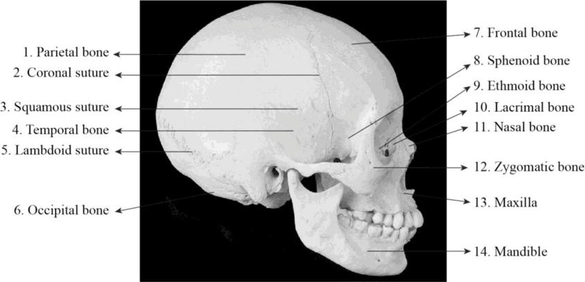
Fig1: Lateral view of the skull.
Explanation of Solution
1. Parietal bone: These are paired cranial bones that constitute the superior lateral sides of the cranial cavity. The term “paries” means “walls”.
2. Coronal suture: It connects the frontal bone and the parietal bones.
3. Squamous suture: The squamous suture connects the temporal bones and the parietal bones.
4. Temporal bone: These are paired cranial bones that constitute the inferior lateral sides of the cranial cavity and contains the ear organs.
5. Lambdoid suture: It is the suture that connects the parietal bones and the occipital bone.
6. Occipital bone: The occipital bone is an unpaired cranial bone that forms the posterior wall and is a part of the cranial cavity floor. The term “occipit” means “atlas”.
7. Frontal bone: It constitutes the anterior portion of the cranial cavity. It is a bowl-shaped, unpaired cranial bone that is situated in the forehead region. It lies anterior to the parietal bones and posterior to the nasal bones.
8. Sphenoid bone: The sphenoid bone is an unpaired cranial bone that is present posterior to the ethmoid bone. The term “sphen” indicates “wedge” and “edios” indicates “form”.
9. Ethmoid bone: The ethmoid bone is a cranial bone and it forms the anterior portion of the cranial cavity floor. The term “ethmos” means “sieve”.
10. Lacrimal bone: The lacrimal bone is a paired facial bone. It is a portion of the eye socket and is present near the nasal bones.
11. Nasal bone: The nasal bones are paired facial bones that form the bridge of the nose.
12. Zygomatic bone: It is also called the cheekbone. It is a paired facial bone. The term “zygoma” indicates “yoke or bar”.
13. Maxilla: The maxilla is otherwise called upper jaw bone. These bones are paired. The maxillae are joined by the intermaxillary suture in the midline and thereby forms the upper jaw.
14. Mandible: The mandible bone otherwise the lower jaw bone is an unpaired facial bone. The term “mandere” indicates “to chew”. It is the only movable bone present in the cranium.
To label: The cranial bones, facial bones, and sutures in figure 9.2.
Answer to Problem 1.1BGL
Pictorial representation: The superior view of the skull is shown in figure 2 given below:
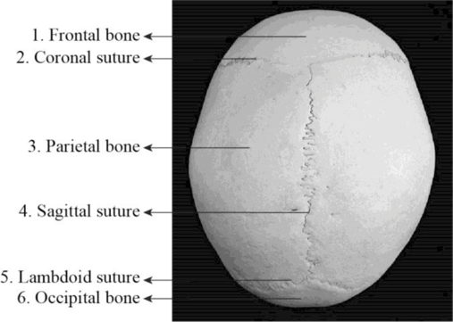
Fig 2: Superior view of the skull.
Explanation of Solution
1. Frontal bone: It constitutes the anterior portion of the cranial cavity. It is an unpaired cranial bone.
2. Coronal suture: The coronal suture connects the frontal bone and the parietal bones.
3. Parietal bone: The paired cranial bones that form the superior lateral walls of the cranial cavity are the parietal bones. Each parietal bone has four margins, four angles, and four surfaces.
4. Sagittal suture: The sagittal suture joins the parietal bones. The term “sagitta” indicates “arrow”.
5. Lambdoid suture: It links the occipital bone with the two parietal bones. The shape of these sutures appears similar to the Greek letter lambda and so it is called lambdoid.
6. Occipital bone: It is an unpaired cranial bone. The term “occipit” means “atlas”. It houses the cerebellum.
To label: The cranial bones, facial bones, and sutures in figure 9.3.
Answer to Problem 1.1BGL
Pictorial representation: The inferior view of the skull is shown in figure 3 given below:
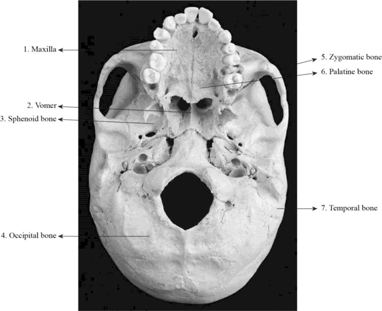
Fig 3: Inferior view of the skull.
Explanation of Solution
1. Maxilla: The maxillae are joined by the intermaxillary suture in the midline and thereby forms the upper jaw. It is a paired facial bone.
2. Vomer: The inferior portion of the nasal septum is formed by the vomer. The vomer is an unpaired facial bone. The term “vomer” indicates “plowshare”. The vomer articulates along with maxilla, ethmoid, palatine, and sphenoid bones.
3. Sphenoid bone: It is an unpaired cranial bone that is present posterior to the ethmoid bone.
4. Occipital bone: It is an unpaired cranial bone that forms the posterior wall and is a part of the cranial cavity floor. The occipital bone articulates along with the cervical spine and also with other bones of the cranium.
5. Zygomatic bone: It is a paired facial bone. It is also called the cheekbone. It articulates with sphenoid bone, frontal bone, temporal bones and the maxilla.
6. Palatine bone: It is a paired facial bone. They form the posterior portion of the hard palate.
7. Temporal bone: These bones are paired cranial bones that form the inferior lateral walls of the cranial cavity and also houses the ear organs.
To label: The cranial bones, facial bones, and sutures in figure 9.4.
Answer to Problem 1.1BGL
Pictorial representation: The superior view of the cranial cavity is shown in figure 4 given below:
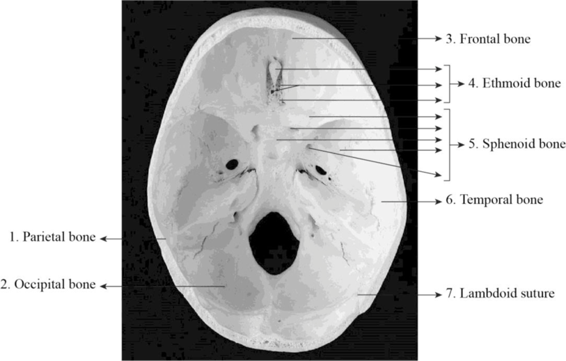
Fig 4: Superior view of cranial cavity floor.
Explanation of Solution
1. Parietal bone: It form the superior lateral walls of the cranial cavity. It is situated on either side of the cranium. They have an irregular quadrilateral shape.
2. Occipital bone: It is an unpaired cranial bone. The bone surface markings of the occipital bone include foramen magnum, occipital condyles, hypoglossal foramina, external occipital protuberance, and others.
3. Frontal bone: It is an unpaired facial bone and it is bowl-shaped. It is situated in the forehead region.
4. Ethmoid bone: It is an unpaired cranial bone. The ethmoid bone consists of cibriform plate, ethmoidal labyrinth, and perpendicular plate.
5. Sphenoid bone: The sphenoid bone is an unpaired cranial bone. The body, pterygoid process, greater wing and the lesser wing are the main parts present in the sphenoid bone.
6. Temporal bone: It is a paired cranial bone. It houses the parts of the inner ear. It is situated at the lateral side of the cranium.
7. Lambdoid suture: The lambdoid is the suture that joins the parietal bones with the occipital bones of the cranium. The lambdoid suture is also known as lambdoidal suture and is found in the posterior region of the cranium.
To label: The cranial bones, facial bones, and sutures in figure 9.5.
Answer to Problem 1.1BGL
Pictorial representation: The anterior view of the skull is shown in figure 5 given below:
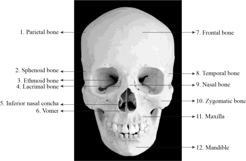
Fig 5: Anterior view of the skull.
Explanation of Solution
1. Parietal bone: It is a paired cranial bone and is located on either side of the cranium. These bones form the roof of the cranium.
2. Sphenoid bone: It is an unpaired cranial bone that is present posterior to the ethmoid bone. It is considered as the most complex bone present in the human body. The term “sphen” indicates “wedge” and “edios” indicates “form”.
3. Ethmoid bone: It is a cranial bone and it is a single, midline bone that forms the anterior portion of the cranial cavity floor. The term “ethmos” means “sieve”.
4. Lacrimal bone: The lacrimal bone is a paired facial bone. It is a portion of the eye socket and is present near the nasal bones.
5. Inferior nasal concha: It is a paired facial bone that forms the posterior lateral walls of the nasal cavity. The term “concha” means “shell”.
6. Vomer: The vomer is a small, thin bone that separates the right and left nasal cavities situated inferior to the perpendicular plate, which forms the superior portion of the nasal septum. It is an unpaired facial bone.
7. Frontal bone: It constitutes the anterior portion of the cranial cavity. It is a bowl-shaped, unpaired cranial bone that is situated in the forehead region. It lies anterior to the parietal bones and posterior to the nasal bones.
8. Temporal bone: The temporal bones are paired cranial bones that form the inferior lateral walls of the cranial cavity and also houses the ear organs.
9. Nasal bone: The nasal bone is a paired facial bone. Their size and form differs in each individual. The nasal bones form the bridge of the nose.
10. Zygomatic bone: The zygomatic bone or cheekbone is a paired facial bone. The zygomatic bone consists of three surfaces, two processes, and four borders.
11. Maxilla: It is a paired facial bone. The maxillae are joined by the intermaxillary suture in the midline and thereby forms the upper jaw. The roots of the upper teeth lie within the alveoli (tooth socket) of the maxillae.
12. Mandible: The mandible bone or the lower jaw bone is an unpaired facial bone. The mandible is the only movable bone present in the cranium. The roots of the lower teeth lie within the alveoli of the mandible.
Want to see more full solutions like this?
Chapter 9 Solutions
Laboratory Manual for Anatomy and Physiology, 6e Loose-Leaf Print Companion


 Human Anatomy & Physiology (11th Edition)BiologyISBN:9780134580999Author:Elaine N. Marieb, Katja N. HoehnPublisher:PEARSON
 Biology 2eBiologyISBN:9781947172517Author:Matthew Douglas, Jung Choi, Mary Ann ClarkPublisher:OpenStax
 Anatomy & PhysiologyBiologyISBN:9781259398629Author:McKinley, Michael P., O'loughlin, Valerie Dean, Bidle, Theresa StouterPublisher:Mcgraw Hill Education,
 Molecular Biology of the Cell (Sixth Edition)BiologyISBN:9780815344322Author:Bruce Alberts, Alexander D. Johnson, Julian Lewis, David Morgan, Martin Raff, Keith Roberts, Peter WalterPublisher:W. W. Norton & Company
 Laboratory Manual For Human Anatomy & PhysiologyBiologyISBN:9781260159363Author:Martin, Terry R., Prentice-craver, CynthiaPublisher:McGraw-Hill Publishing Co.
 Inquiry Into Life (16th Edition)BiologyISBN:9781260231700Author:Sylvia S. Mader, Michael WindelspechtPublisher:McGraw Hill Education