
Concept explainers
Videos
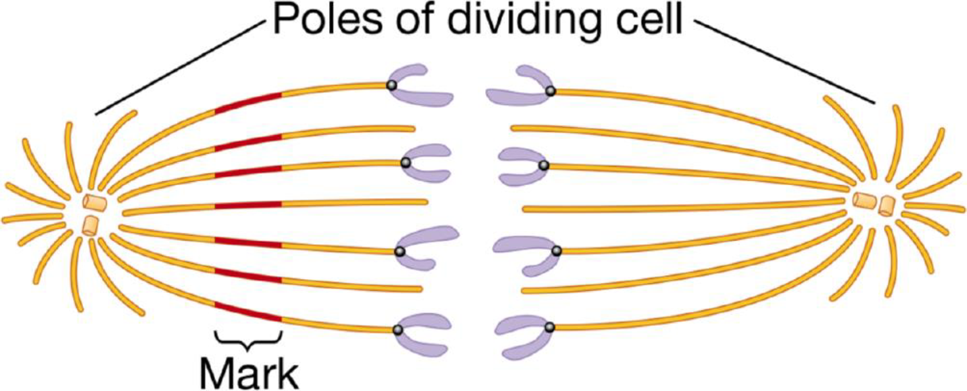
SCIENTIFIC THINKING Microtubules often produce movement through their interaction with motor proteins. But in some cases, microtubules move cell components when the length of the microtubule changes. Through a series of experiments, researchers determined that microtubules grow and shorten as tubulin proteins are added or removed from their ends. Other experiments showed that microtubules make up the spindle apparatus that “pulls” chromosomes toward opposite ends (poles) of a dividing cell. The figures below describe a clever experiment done in 1987 to determine whether a spindle microtubule shortens (depolymerizes) at the end holding a chromosome or at the pole end of a dividing cell.
Experimenters labeled the microtubules of a dividing cell from a pig kidney with a yellow fluorescent dye. As shown on the left half of the diagram below, they then marked a region halfway along the microtubules by using a laser to eliminate the fluorescence from that region. They did not mark the other side of the spindle (right side of the figure).
The figure below illustrates the results they observed as the chromosomes moved toward the opposite poles of the cell.
The figure below illustrates the results they observed as the chromosomes moved toward the opposite poles of the cell.
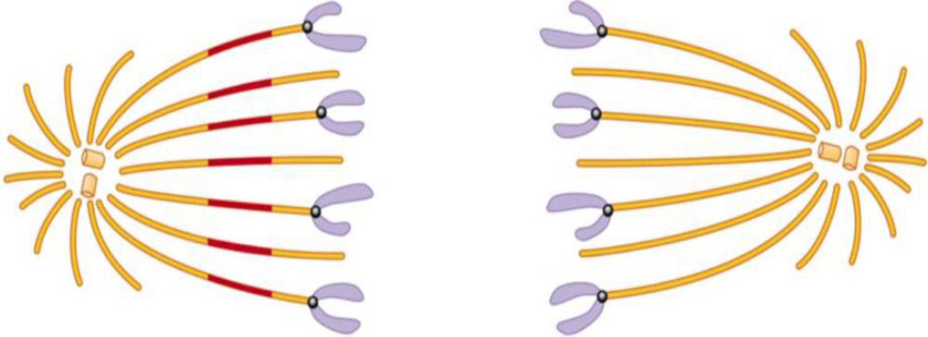
Describe these results. What would you conclude about where the microtubules depolymerize from comparing the length of the microtubules on either side of the mark? How could the experimenters determine whether this is the mechanism of chromosome movement in all cells?
Want to see the full answer?
Check out a sample textbook solution
Chapter 4 Solutions
CAMPBEL BIOLOGY:CONCEPTS & CONNECTIONS


- The drug vinblastine is used in cancer therapy to stop the runaway division of cancer cells. Vinblastine inhibits the assembly and growth of microtubules. Explain how the action of this drug prevents mitosis (refer to Figure 2.17).arrow_forwardThe effect of MAP2 on microtubules is similar to the effect of CapZ on microfilaments how? both stabilize the positive end of the filament both bundle the filaments in parallel bundles both increase the instability of the negative end of the filament both nucleate the formation of the filamentsarrow_forwardScientists often extract DNA from the nucleus of cells for analysis or use. This process breaks the cell membrane, spins the solution to remove the large particles through high g-force, adds alcohol to make the DNA less soluble aqueous solution, then spins the solution again to pull the DNA out of solution. Consider a cell in metaphase compared to a cell in rest (not in the cell cycle leading to cell division). What properties of a metaphase cell might let you extract more DNA compared to the resting cell? Are there any that might make the extraction more difficult?arrow_forward
- Match each of the following to its role in the cell. Microtubules Actin Tubulin Intermediate filaments Kinesin Dynein Myosin Centrosome A. Cytoskeletal fiber that is intermediate in diameter and very sturdy. Does not form from polymerization of protein monomers like the other ones we discussed in class, but consists of polypeptide strands. Keratin and Lamin A are examples. B. The structure that initiates polymerization of new microtubules and anchors microtubules in the center of the cell near the Golgi Complex. C. Motor protein whose activity results in contraction of muscle. D. Thinnest cytoskeletal filament. Responsible for initiation of amoeboid cell movement via pseudopod formation. E. The "monomer" unit of the thickest cytoskeletal polymer; a dimer of two polypeptide subunits. F. Cytoskeletal structure with the…arrow_forwardThe drug taxol is extracted from the bark of yew trees. It binds tightly to microtubules and stabilizes them. When added to cells, it causes much of the free tubulin to assemble into microtubules. Taxol can be used as an anticancer drug. At the molecular level, what does the drug prevent? At the cellular level, what part of cell division does it prevent? Be specific.arrow_forwardScientists in a lab have isolated a substance that prevents cells from synthesizing spindle fibres. How would this substance affect cell division? Explain.arrow_forward
- Consider the proper progression of the cell cycle. In one of the first steps, the __ Select one: a. cell membrane breaks down as the micrtubulin filaments dissolve b. cell membrane breaks down as the intermediate filaments dissolve c. nuclear membrane breaks down as the actin filaments dissolve d. cell membrane breaks down as the actin filaments dissolve e. nuclear membrane breaks down as the intermediate filaments dissolvearrow_forwardIntegrins are important in cell crawling because they anchor the leading edge of the cell to the surface it is moving over they are receptors for diffusible chemical ligands that determine the direction the cell will move in they cross-link actin filaments to microtubules and intermediate filaments of the cytoskeleton they serve as nucleating sites for polymerization of new actin filamentsarrow_forwardIn cancer, cells divide out of control. Thus, chemicals that interfere with cell division might be used to stop cancer cells. How many of the following might stop human cancer cells? I. A chemical that prevents the phosphorylation of lamins. II. A chemical that prevents microtubule shortening. III. A chemical that prevents actin-myosin contraction. IV. A chemical that prevents the dephosphorylation of lamins. O 2 O 4 O 1arrow_forward
- An antibody that binds to myosin prevents the movement of myosin molecules along actin filaments. What is the main role of an antibody? What would be the effect of injecting this antibody into cells on the movement of chromosomes at anaphase? Explain your answer. What would be the effect of injecting this antibody into cells on cytokinesis? Explain the result.arrow_forwardBiologists have long been interested in the effects of radiation on cells. In one experiment, researchers examined the effect of radium on mitosis of chick embryo cells growing in culture. A population of experimental cells was examined under the microscope for the number of cells in telophase (as a measure of mitosis occurring) before, during, and after exposure to radium. The results are shown in the Figure. What is the effect of radium exposure on mitosis? Source: R. G. Canti and M. Donaldson. 1926. The effect of radium on mitosis in vitro. Proceedings of the Royal Society of London, Series B, Containing Papers of a Biological Character 100:413419.arrow_forwardThe eukaryotic cell in the photo on the left is in the process of cytoplasmic division. Is this cell from a plant or an animal? How do you know?arrow_forward
 Biology: The Dynamic Science (MindTap Course List)BiologyISBN:9781305389892Author:Peter J. Russell, Paul E. Hertz, Beverly McMillanPublisher:Cengage Learning