
Cardiopulmonary Anatomy & Physiology
7th Edition
ISBN: 9781337794909
Author: Des Jardins, Terry.
Publisher: Cengage Learning,
expand_more
expand_more
format_list_bulleted
Question
Chapter 14, Problem 8RQ
Summary Introduction
To review:
The given blank space in the statement using the following ECG graph.
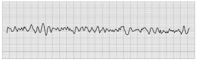
QRS duration: __________ QT duration: ____________
Ventricular rate and rhythm: _____________
Atrial rate rhythm: _____________________
PR interval: __________________________
Interpretation: ________________________
Introduction:
The diagnostic test machine that is used to monitor a patient’s heart rate is known as Electrocardiogram (ECG). The ECG records the electrical signals, which are in the form of wave components, such as P, Q, R, S, T, and U. The doctors interpret this ECG through a systemic approach.
Expert Solution & Answer
Want to see the full answer?
Check out a sample textbook solution
Students have asked these similar questions
The standard EKG consists of 10 sensors that record 12 leads of the heart’s electrical activity from different angles, allowing for a thorough three-dimensional interpretation of its activity. This is transmitted by the electrodes to the equipment to be interpreted and is used to diagnose cardiac medical conditions. In case of an abnormal EKG, the second step would be to use a Holter monitor.
How would you explain how to perform an EKG (steps)?
Where will you place the electrodes when performing and EKG? Why?
What are the different lead types, connections, and placements?
When you conclude an EKG, what are the different components that you need to observe and confirm before you disconnect the patient? Can you explain the difference between normal, abnormal, and artifacts?
What is a Holter monitor? Under what circumstances would one be ordered for a patient?
How do you use a Holter monitor?
Educate a patient: What you will do before, during, and after an electrocardiogram or…
Below is an image of an electrocardiogram (EKG) tracing. What does "4" represent?
2
3
1
дел
4
P
S
T
R
nh
LO
5
Electrocardiogram is also called ECG. It is a test that assesses the electrical activity of the heart through electrodes attached to the skin. The figure (A) on the X axis - represents the time and on the Y axis - the mV of an electrocardiogram. Image B represents the electrocardiogram of a rested adult patient. Based on the images, identify and comment on the diagnosis.
Chapter 14 Solutions
Cardiopulmonary Anatomy & Physiology
Knowledge Booster

Similar questions
- One of the following is a life-threatening arrhythmia and an absolute emergency that should immediately be notified by the MA to the physician in the event it appears in the EKG strips. One of the following is a life-threatening arrhythmia and an absolute emergency that should immediately be notified by the MA to the physician in the event it appears in the EKG strips. Sinus rhythm Sinus tachycardia Sinus bradycardia Ventricular fibrillationarrow_forwardIt has been agreed a long time ago that the schematics of cardiovascular system will be color coded - the _____________________________________ are depicted in red, while ___________________________________ are drawn in blue. In this image, A is (specific vessel) _____________________________________________________. B is (specific vessel)arrow_forwardMark the following coordinates on the image about: 1. repolarization of the ventricles 2. conduction through the atrioventricular node, 3. depolarization of the ventricles and repolarization of the atria. 4. First deflection corresponding to the movement of current when the atria depolarizearrow_forward
- Analyze this VVI pacemaker strip. Identify any underlying rhythm and how the pacemaker is functioning. This rhythm strip shows atrial fibrillation with... WI pacing with intermittent failure to sense WI pacing with intermittent failure to capture Normal WVI pacemaker function WI pacing with intermittent failure to sense and failure to capture WI pacing with oversensing TOOLS INSTRUCTIONS If needed, select one of the above tools. SUE ITarrow_forwardWhat are echocardiogram, holter monitoring, and the electrocardiogram. what information we can gain from the tests and any limitations to the tests.arrow_forwardA 67-year-old-man is in the operating room undergoing a hip replacement. The procedure is going along uneventfully, and there is no indication of acute blood loss. Suddenly, the patient develops supraventricular tachycardia. Intravenous injection is administered and within 15 s, the electrocardiogram shows normal sinus rhythm. What is the most likely drug responsible for this normalization of the electrocardiogram? Select one: O ignocaine Quinidine Esmolol Verapamil Adenosinearrow_forward
- Analyze this rhythm strip and identify the rhythm/dysrhythmia. Sinus rhythm with bundle branch block Second-degree AV block type II Second-degree AV block type ! Sinus rhythm with first-degree AV block Third-degree AV block 2:1 second-degree AV block SUBMIT INSTRUCTIONS If needed, select one of the above tools. بلس السل سلسالس السلسلةarrow_forwardPlease do your own solution and dont copy paste from other posters answers. Draw a basic block diagram of oscillometric blood Pressure measurement setup and explain each block in the diagram in details.arrow_forwardNumber the following structures of the cardiac conduction system in the normal order of depolarization (1 to 5). _______________ AV bundle (of His) _______________ AV node _______________ Purkinje (conduction) fibers _______________ Right and left bundle branches _______________ SA nodearrow_forward
- What major difference(s) did you observe between the right and left ventricle? ____________________________________________________________________________________arrow_forwardThe standard EKG consists of 10 sensors that record 12 leads of the heart’s electrical activity from different angles, allowing for a thorough three-dimensional interpretation of its activity. This is transmitted by the electrodes to the equipment to be interpreted and is used to diagnose cardiac medical conditions. In case of an abnormal EKG, the second step would be to use a Holter monitor. How would you explain to your classmates how to perform an EKG (steps)? Where will you place the electrodes when performing and EKG? Why? What are the different lead types, connections, and placements? When you conclude an EKG, what are the different components that you need to observe and confirm before you disconnect the patient? Can you explain the difference between normal, abnormal, and artifacts? What is a Holter monitor? Under what circumstances would one be ordered for a patient? How do you use a Holter monitor? Educate a patient: What you will do before, during, and after an…arrow_forwardYour research participant, Bryan, comes in for his cardiopulmonary exercise test (CPET). You want to run a VO2 max and EKG on him using the Bruce protocol. During the initial review of medical history, Bryan mentions he has been having some angina symptoms all day. On the angina scale he rated this as a 3. Would you test this person, yes or no? If yes, please indicate what protocol you would choose and whether or not the test would be maximal or submaximal? Please include your rationale.arrow_forward
arrow_back_ios
SEE MORE QUESTIONS
arrow_forward_ios
Recommended textbooks for you
 Cardiopulmonary Anatomy & PhysiologyBiologyISBN:9781337794909Author:Des Jardins, Terry.Publisher:Cengage Learning,
 Understanding Health Insurance: A Guide to Billin...Health & NutritionISBN:9781337679480Author:GREENPublisher:Cengage
 Human Physiology: From Cells to Systems (MindTap ...BiologyISBN:9781285866932Author:Lauralee SherwoodPublisher:Cengage Learning
Cardiopulmonary Anatomy & Physiology
Biology
ISBN:9781337794909
Author:Des Jardins, Terry.
Publisher:Cengage Learning,
Understanding Health Insurance: A Guide to Billin...
Health & Nutrition
ISBN:9781337679480
Author:GREEN
Publisher:Cengage
Human Physiology: From Cells to Systems (MindTap ...
Biology
ISBN:9781285866932
Author:Lauralee Sherwood
Publisher:Cengage Learning