
Concept explainers
To review:
The given blank space in the statement using the following ECG graph.
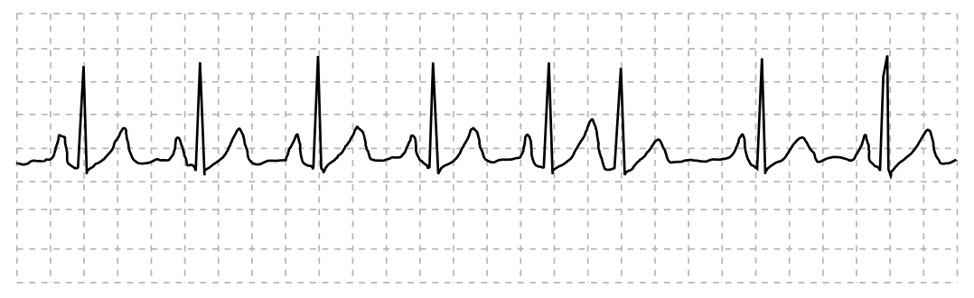
QRS duration: __________ QT duration: ____________
Ventricular rate and rhythm: _____________
Atrial rate rhythm: _____________________
PR interval: __________________________
Interpretation: ________________________
Introduction:
The electrocardiogram or ECG is a tool that is used to record a patient’s heart rate in real time. The heartbeat produces electrical signals that are recorded in this tool. It is recorded in a waveform, which has P, Q, R, S, T, and U components. The ECG is interpreted by doctors to know the condition of patients.
Explanation of Solution
The heart rate of the patient is regular with normal electrical activity of the heart. There was one PAC (premature atrial contractions), also known as atrial premature beats, at sixth position, which occurred naturally. The ECG tracing is interpreted as normal sinus rhythm.
QRS duration: The duration of the QRS complex is less than 0.12 seconds.
QT duration: It is more than 0.12 seconds.
Ventricular rate and rhythm: The ventricular rate is regular and between 60 and 100bpm. Ventricular rhythm is less than 0.12 seconds.
Atrial rate rhythm: It is regular with the heart rate up to 100 bpm and occurrence of one PAC at the 6th complex.
PR interval: Duration is less than 0.20 seconds.
Interpretation: The patient’s heart rate is between 60 and 100 bpm, so the graph represents ECG tracing of Normal Sinus Rhythm (NSR) with one PAC at the sixth complex.
QRS duration: 0.6 seconds QT duration: 0.40 seconds
Ventricular rate and rhythm: 86 bpm
Atrial rate rhythm: 86 bpm
PR interval: 0.16 seconds
Interpretation: The graph shows normal sinus rhythm with 86 bpm having one atrial premature beat at the sixth complex
Want to see more full solutions like this?
Chapter 14 Solutions
Cardiopulmonary Anatomy & Physiology


- Fill information below on identifying the ekg rhythm striparrow_forwardSelect the correct answer for each question. Explain why you chose that answer as opposed to the others. In other words, explain why the other choices are incorrect. Select the correct statement about cardiac output. A slow heart rate increases end diastolic volume, stroke volume, and force of contraction. Decreased venous return will result in increased end diastolic volume. Stroke volume increases if end diastolic volume decreases. If a semilunar valve were partially obstructed the end systolic volume in the affected ventricle would be decreased.arrow_forwardThe four-chambered heart is a single organ but is sometimes described as a “double pump” because it functions more like two pumps than one. Explain why this is so, including the names of the heart structures involved. please helparrow_forward
- (a) Referring to the figure, find the time systolic pressure lags behind the middle of the QRS complex. (b) Discuss the reasons for the time lag.arrow_forwardIn the image attached help me locate the anterior ventricular artery and draw a thrombosis in that artery and then shade the area of the heart that would experience a myocardial infarction. Thanksarrow_forwardMark the following coordinates on the image about: 1. repolarization of the ventricles 2. conduction through the atrioventricular node, 3. depolarization of the ventricles and repolarization of the atria. 4. First deflection corresponding to the movement of current when the atria depolarizearrow_forward
- If Gerald's heart rate was 72 beats per minute, determine the number of QRS complexes that would have appeared on an EKG during the first 30 seconds.arrow_forwardDuring which phase of the cardiac cycle is the ST segment present on an EKG? Group of answer choices Atrial systole Isovolumetric ventricular contraction Rapid ventricular ejection Isovolumetric ventricular relaxation Rapid ventricular fillingarrow_forwardIn electrocardiography (=EKG, =ECG), what causes a P wave?______ A. atrial repolarization B. ventricular repolarization C. atrial depolarization D. ventricular depolarizationarrow_forward
- HQ4. Label the posterior view of the heart.arrow_forwardcardiac surgery requires cardiac instruments and ___________.arrow_forwardLook at the answer and give the reason for answer. Write why the answer is the answer. Q 6 - What are the implications for ventricular filling? Answer: The ventricles will fill with less blood. Q 7 - In a healthy heart, which of the following serves as pacemaker for the conduction system? Answer: SA node Q 8 - Based on information provided in this chapter, what happens to the cardiac output of a patient following an MI? Answer: The cardiac output is decreased. Q 9 - Based on information provided in this chapter, what are the main goals of drug therapy in patients following MI? Answer: decreasing the afterload and decreasing the end systolic ventricular volumearrow_forward
 Cardiopulmonary Anatomy & PhysiologyBiologyISBN:9781337794909Author:Des Jardins, Terry.Publisher:Cengage Learning,
 Understanding Health Insurance: A Guide to Billin...Health & NutritionISBN:9781337679480Author:GREENPublisher:Cengage