
Laboratory Manual For Human Anatomy & Physiology
4th Edition
ISBN: 9781260159363
Author: Martin, Terry R., Prentice-craver, Cynthia
Publisher: McGraw-Hill Publishing Co.
expand_more
expand_more
format_list_bulleted
Videos
Textbook Question
Chapter 44, Problem F44.15A
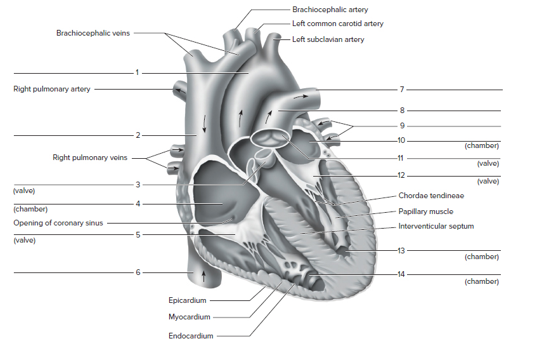
FIGURE 44.15 Label this frontal section of the human heart. The arrows indicate the direction of blood flow.
Expert Solution & Answer
Want to see the full answer?
Check out a sample textbook solution
Students have asked these similar questions
Draw the posterior view of the heart exposing the coronary vessels
In this view of the superior surface of the heart, at the base where all the blood vessels enter and leave the heart, #17 is the opening where the______ enter and transport blood into the _______. (specific chamber)
NOTES
and the left side serving the systemic circuit. In Figure 16-15, complete
the schematic showing the blood flow to and from the heart (the starting
points are given to you). Use a blue pen or pencil to denote the direction of
deoxygenated blood and a red pen or pencil for oxygenated blood flow.
Include the names of the major vessels, chambers, and valves involved,
based on the following list:
lung capillary beds
body capillary beds
right ventricle
left ventricle
bicuspid valve
superior vena cava
tricuspid valve
inferior vena cava
pulmonary semilunar valve
pulmonary trunk
aortic semilunar valve
R. and L. pulmonary arteries
R. and L. pulmonary veins
aorta
Pulmonary Circulation
Systemic Circulation
Right atrium
Left atrium
URA--YCK
HAT
Lungs
Body
Figure 16-15. Schematic of circulation
Chapter 44 Solutions
Laboratory Manual For Human Anatomy & Physiology
Ch. 44 - The ________is the inferior end of the heart that...Ch. 44 - Oxygen-rich blood is located in the a. left-side...Ch. 44 - The two superior heart chambers are the a. left...Ch. 44 - Which of the following is an atrioventricular (AV)...Ch. 44 - The __________ lines the heart chambers. a....Ch. 44 - Which heart valve has two cusp s instead of three...Ch. 44 - Chordae tendineae connect the cusps of the AV...Ch. 44 - The systemic circuit delivers blood to the lungs...Ch. 44 - The right and left coronary arteries containing...Ch. 44 - FIGURE 44.13 Identity the features on this...
Ch. 44 - FIGURE 44.14 identity the features indicated on...Ch. 44 - FIGURE 44.15 Label this frontal section of the...Ch. 44 - Prob. 2.1ACh. 44 - Compare the structure of the left atrioventricular...Ch. 44 - Prob. 3.2ACh. 44 - What is the functional significance of the...Ch. 44 - List the correct pathway through which blood must...Ch. 44 - Describe the general overall shape of the left and...Ch. 44 - What was the measured thickness of the left...Ch. 44 - Explain the functional significance of the...
Knowledge Booster


Learn more about
Need a deep-dive on the concept behind this application? Look no further. Learn more about this topic, biology and related others by exploring similar questions and additional content below.Similar questions
- Could you draw a picture of the flow of blood through the heart. And label at least 10 chambers,vessels, or structures. Could you also in the drawing include colors indicate which chambers or vessels are carrying oxygenated (red) blood or deoxygenated blood (blue)arrow_forwardtrace a drop of blood from the left ventricle of the heart to the wrist of the right hand and back to the heart(name each vessel) then trace the drop of blood to the dorsum of the right foot and back to the right side of the heart.arrow_forwardThe heart is lateral to the lungs. True or falsearrow_forward
- Structure of the Heart Use this table as a checklist for your study of the heart. Do not forget to fill in the function column. Structure Right atrium Computer Simulation Function(s) Sheep Human Left atrium Right ventricle ロ Left ventricle Interventricular sulcus Anterior interventricular artery ロ Great cardiac vein Small cardiac vein Right coronary artery Circumflex artery Left coronary artery Aorta Pulmonary artery Superior vena cava Inferior vena cava Interventricular septum Myocardium ロ Epicardium Parietal pericardium Pericardial space Fibrous pericardium ロ Mitral valve Tricuspid valve Chordae tendineae Papillary muscle ロ Aortic semilunar valve Pulmonary semilunar valve Copyright 2003 by Mosby, Inc. All rights reserved. 323arrow_forwardIn the image attached help me locate the anterior ventricular artery and draw a thrombosis in that artery and then shade the area of the heart that would experience a myocardial infarction. Thanksarrow_forwardOn a diagram of a frontally sectioned heart, indicate the location of the cardiac skeleton.arrow_forward
- fill in the blanks Blood that has circulated to the heart muscle itself also returns to the right atrium. Once blood leaves the cells of the myocardium, it flows into a system of cardiac veins, eventually flowing into the great cardiac vein, and then the coronary sinus. On this anterior view of the heart, locate the great cardiac vein and the coronary sinus (shown through to the back). Slide 14: Identify the two large veins that merge and flow into the superior vena cava: A._________________________________ and B._________________________________. (Note that unlike the arterial system, there are two veins, a right and a left, with this name.) Locate either the right or left brachiocephalic vein. Each is formed by the union of two slightly smaller veins. An C.___________________________ vein draining the head and a D.______________________ vein bringing blood back from the arm. Locate an additional vein draining blood from the head that flows into the subclavian vein, the…arrow_forwardLabel the three views of the heart in Figure 12.1 with the following O Right atrium O Left atrium Great Vessels O Superior vena cava O Inferior vena cava Structures of the Ventricles O Right ventricle O Left ventricle O Interventricular septum O Chordae tendineae O Papillary muscles O Pulmonary trunk O Pulmonary veins O Aorta Coronary Vessels O Right coronary artery O Anterior interventricular artery (left anterior descending) O Coronary sinus O Great cardiac vein O Circumflex artery Atrioventricular Valves O Tricuspid valve O Mitral valve Semilunar Valves O Pulmonary valve O Aortic valve Carrow_forwardLabel the three views of the heart in Figure 12.1 with the following terms. O Right atrium O Left atrium Great Vessels O Superior vena cava O Inferior vena cava Structures of the Ventricles O Right ventricle O Left ventricle O Interventricular septum O Chordae tendineae O Papillary muscles O Pulmonary trunk O Pulmonary veins O Aorta Coronary Vessels O Right coronary artery O Anterior interventricular artery (left anterior descending) O Coronary sinus O Great cardiac vein O Circumflex artery Atrioventricular Valves O Tricuspid valve O Mitral valve Semilunar Valves O Pulmonary valve O Aortic valve B FIGURE 12.1 Heart: (A) anterior view; (B) frontal section; (C) posterior viewarrow_forward
- Consider the circulation of blood through the heart. Put the structures in the order that blood flows through them, starting with the superior and inferior venae cavae|(#1) and ending with the ascending aorta (#14).arrow_forwardIn a paragraph or using an arrows, trace the blood flow from the superior vena cava, inferior vena cava and coronary sinus entering the heart to the pulmonary circulation and systemic circulation.arrow_forwardIn the figure below on the left, label the P, QRS and T waves. Describe what is happening in the heart in the P wave: What is occurring in the electrical conduction system? Circle in the figure on the right, what part (if any) of the electrical conduction system is activated.arrow_forward
arrow_back_ios
SEE MORE QUESTIONS
arrow_forward_ios
Recommended textbooks for you
 Human Physiology: From Cells to Systems (MindTap ...BiologyISBN:9781285866932Author:Lauralee SherwoodPublisher:Cengage Learning
 Biology 2eBiologyISBN:9781947172517Author:Matthew Douglas, Jung Choi, Mary Ann ClarkPublisher:OpenStax
Human Physiology: From Cells to Systems (MindTap ...
Biology
ISBN:9781285866932
Author:Lauralee Sherwood
Publisher:Cengage Learning
Biology 2e
Biology
ISBN:9781947172517
Author:Matthew Douglas, Jung Choi, Mary Ann Clark
Publisher:OpenStax
Respiratory System; Author: Amoeba Sisters;https://www.youtube.com/watch?v=v_j-LD2YEqg;License: Standard youtube license