
Laboratory Manual For Human Anatomy & Physiology
4th Edition
ISBN: 9781260159363
Author: Martin, Terry R., Prentice-craver, Cynthia
Publisher: McGraw-Hill Publishing Co.
expand_more
expand_more
format_list_bulleted
Videos
Textbook Question
Chapter 44, Problem F44.14A
FIGURE 44.14 identity the features indicated on this anterior view of a frontal section of a human heart model using the terms provided (Note: The pulmonary valve is not shown on the portion of the model photographed)
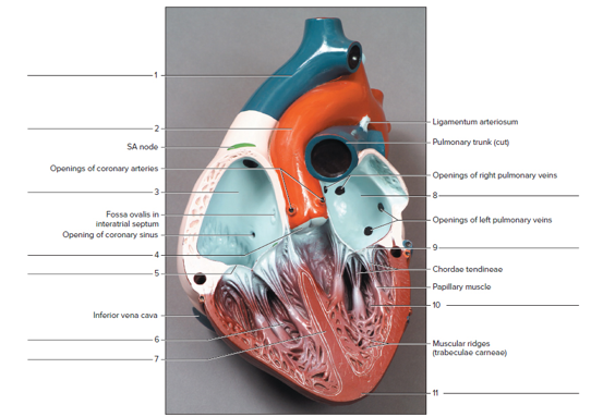
Terms:
Aorta Left atrium
Right ventricle
Aortic valve
Left ventricle
Superior vena cava
Apex of heart
Mitral valve
Tricuspid valve
Interventricularseptum
Right atrium
Expert Solution & Answer
Want to see the full answer?
Check out a sample textbook solution
Students have asked these similar questions
Structure of the Heart
Use this table as a checklist for your study of the heart. Do not forget to fill in the function column.
Structure
Right atrium
Computer
Simulation Function(s)
Sheep
Human
Left atrium
Right ventricle
ロ
Left ventricle
Interventricular sulcus
Anterior interventricular artery
ロ
Great cardiac vein
Small cardiac vein
Right coronary artery
Circumflex artery
Left coronary artery
Aorta
Pulmonary artery
Superior vena cava
Inferior vena cava
Interventricular septum
Myocardium
ロ
Epicardium
Parietal pericardium
Pericardial space
Fibrous pericardium
ロ
Mitral valve
Tricuspid valve
Chordae tendineae
Papillary muscle
ロ
Aortic semilunar valve
Pulmonary semilunar valve
Copyright 2003 by Mosby, Inc. All rights reserved.
323
Drag the steps below and place them in the order of blood flow through the heart.
First:
Second:
Third:
Fourth:
Fifth:
Sixth:
Seventh:
Eighth:
Ninth:
Tenth:
Flows through right atrial ventricular valve (tricuspid)
Blood back flowing in aorta toward heart enters coronary circulation
Flowing into the right atrium
Out the aortic semilunar valve into the aorta
Back to heart through the pulmonary veins to the left atrium
Into the right ventricle
Into the left ventricle
Left atrium through left atrial ventricular valve (BICUSPID OR MITRAL)
Out the pulmonary semilunar valve through the pulmonary arteries to lungs
To the body through the aortic branches
NOTES
and the left side serving the systemic circuit. In Figure 16-15, complete
the schematic showing the blood flow to and from the heart (the starting
points are given to you). Use a blue pen or pencil to denote the direction of
deoxygenated blood and a red pen or pencil for oxygenated blood flow.
Include the names of the major vessels, chambers, and valves involved,
based on the following list:
lung capillary beds
body capillary beds
right ventricle
left ventricle
bicuspid valve
superior vena cava
tricuspid valve
inferior vena cava
pulmonary semilunar valve
pulmonary trunk
aortic semilunar valve
R. and L. pulmonary arteries
R. and L. pulmonary veins
aorta
Pulmonary Circulation
Systemic Circulation
Right atrium
Left atrium
URA--YCK
HAT
Lungs
Body
Figure 16-15. Schematic of circulation
Chapter 44 Solutions
Laboratory Manual For Human Anatomy & Physiology
Ch. 44 - The ________is the inferior end of the heart that...Ch. 44 - Oxygen-rich blood is located in the a. left-side...Ch. 44 - The two superior heart chambers are the a. left...Ch. 44 - Which of the following is an atrioventricular (AV)...Ch. 44 - The __________ lines the heart chambers. a....Ch. 44 - Which heart valve has two cusp s instead of three...Ch. 44 - Chordae tendineae connect the cusps of the AV...Ch. 44 - The systemic circuit delivers blood to the lungs...Ch. 44 - The right and left coronary arteries containing...Ch. 44 - FIGURE 44.13 Identity the features on this...
Ch. 44 - FIGURE 44.14 identity the features indicated on...Ch. 44 - FIGURE 44.15 Label this frontal section of the...Ch. 44 - Prob. 2.1ACh. 44 - Compare the structure of the left atrioventricular...Ch. 44 - Prob. 3.2ACh. 44 - What is the functional significance of the...Ch. 44 - List the correct pathway through which blood must...Ch. 44 - Describe the general overall shape of the left and...Ch. 44 - What was the measured thickness of the left...Ch. 44 - Explain the functional significance of the...
Knowledge Booster


Learn more about
Need a deep-dive on the concept behind this application? Look no further. Learn more about this topic, biology and related others by exploring similar questions and additional content below.Similar questions
- What is the tissue type for the following structures of the heart Fibrous pericardium Serous pericardium Myocardium Endocardium Right atrium right Ventricle Left Atrium Left Ventricle Auricles Papillary muscles Fossa Ovalis Pectinate Muscles Chordae Tendinae Interventricular septum Tricuspid, pulmonary, Bicuspid, mitral, and Aortic semilunar valves pulmonary trunk Pulmonary artery Aorta Arteries Sinus/veinsarrow_forwardIdentify whether the given statement is true or false -Left ventricle->Left atrium->Right ventricle->Right atrium is the correct sequence for blood entering the heart through the vena cavae and leaving through the aorta. -The sinoatrial node is situated in the wall of the interventricular septum. -Impulses through the conduction system of the heart follow the ordered path: SA node->conduction myofibers->AV bundle->AV node. -The heart is covered by the suprardium. -The “lub” sound of the heart is caused by closing of the semilunar valves. -To clearly hear the heart sound of the bicuspid valve, a stethoscope should be placed to the left of the sternum at the second intercostal space. -The left ventricular wall of the heart is thicker than the right wall in order to pump blood with a greater pressure. -When the atrioventricular bundle is completely interrupted the ventricles typically contract at 30 to 40 beats/min. -At late diastole, the atria and ventricles are relaxed…arrow_forwardIn the image attached help me locate the anterior ventricular artery and draw a thrombosis in that artery and then shade the area of the heart that would experience a myocardial infarction. Thanksarrow_forward
- Name a structure that is on the ventural side of the heart located more laterally within the axial regionarrow_forwardLabel the three views of the heart in Figure 12.1 with the following O Right atrium O Left atrium Great Vessels O Superior vena cava O Inferior vena cava Structures of the Ventricles O Right ventricle O Left ventricle O Interventricular septum O Chordae tendineae O Papillary muscles O Pulmonary trunk O Pulmonary veins O Aorta Coronary Vessels O Right coronary artery O Anterior interventricular artery (left anterior descending) O Coronary sinus O Great cardiac vein O Circumflex artery Atrioventricular Valves O Tricuspid valve O Mitral valve Semilunar Valves O Pulmonary valve O Aortic valve Carrow_forwardIn the given table, three of the anatomical and physiological terms are similar or related; one does not belong with the other three. Choose the term that does NOT belong in each of the following groups. A B C D 1 Pulmonary Trunk Vena Cava Right Side of the Heart Left Side of the Heart 2 QRS Wave T Wave P Wave Electrical Activity of the Ventricles 3 AV Valves Closed AV Valves Opened Ventricular Systole Semilunar Valves Open 4 Tricuspid Valve Mitral Valve Bicuspid Valve Left AV Valve 5 Pulmonary Valve Umbilical Artery Pulmonary Vein Superior Vena Cavaarrow_forward
- In the figure below on the left, label the P, QRS and T waves. Describe what is happening in the heart in the P wave: What is occurring in the electrical conduction system? Circle in the figure on the right, what part (if any) of the electrical conduction system is activated.arrow_forwardLabel the three views of the heart in Figure 12.1 with the following terms. O Right atrium O Left atrium Great Vessels O Superior vena cava O Inferior vena cava Structures of the Ventricles O Right ventricle O Left ventricle O Interventricular septum O Chordae tendineae O Papillary muscles O Pulmonary trunk O Pulmonary veins O Aorta Coronary Vessels O Right coronary artery O Anterior interventricular artery (left anterior descending) O Coronary sinus O Great cardiac vein O Circumflex artery Atrioventricular Valves O Tricuspid valve O Mitral valve Semilunar Valves O Pulmonary valve O Aortic valve B FIGURE 12.1 Heart: (A) anterior view; (B) frontal section; (C) posterior viewarrow_forwardIdentify whether the following statement is either true or false. Left ventricle->Left atrium->Right ventricle->Right atrium is the correct sequence for blood entering the heart through the vena cavae and leaving through the aorta. The sinoatrial node is situated in the wall of the interventricular septum. Impulses through the conduction system of the heart follow the ordered path: SA node->conduction myofibers->AV bundle->AV node. To clearly hear the heart sound of the bicuspid valve, a stethoscope should be placed to the left of the sternum at the second intercostal space. At late diastole, the atria and ventricles are relaxed and the aortic semilunar valve is open. During ventricular contraction all the blood is forced out of the ventricles.arrow_forward
- In this view of the superior surface of the heart, at the base where all the blood vessels enter and leave the heart, #17 is the opening where the______ enter and transport blood into the _______. (specific chamber)arrow_forwardThe events of the cardiac cycle cause cyclical changes in left ventricular pressure and volume over time. Another way to represent these events is with a pressure-volume loop, as shown below. Drag the labels from the left into the appropriate boxes on the pressure- volume loop to demonstrate your understanding of the cardiac cycle. Aortic valve closure AV valve opening Systolic pressure Isovolumetric relaxation Isovolumetric contraction 120 Diastolic pressure Ventricular filling 80 End-diastolic volume Ventricular ejection 40 AV valve closure End-systolic volume Aortic valve opening 60 120 LV volume (mL) O McGraw-Hill Education Reset LV pressure (mm Hg)arrow_forwardPlease explain blood flow through your body in a flow diagram format (do not put a picture of heart or write a paragraph). Start and end at the inferior/superior vena cava. (HINT: make sure you mention all the valves within the heart and where does blood go from deoxygenated to oxygenated and vice versa).arrow_forward
arrow_back_ios
SEE MORE QUESTIONS
arrow_forward_ios
Recommended textbooks for you
 Biology 2eBiologyISBN:9781947172517Author:Matthew Douglas, Jung Choi, Mary Ann ClarkPublisher:OpenStax
 Human Physiology: From Cells to Systems (MindTap ...BiologyISBN:9781285866932Author:Lauralee SherwoodPublisher:Cengage Learning
Biology 2e
Biology
ISBN:9781947172517
Author:Matthew Douglas, Jung Choi, Mary Ann Clark
Publisher:OpenStax
Human Physiology: From Cells to Systems (MindTap ...
Biology
ISBN:9781285866932
Author:Lauralee Sherwood
Publisher:Cengage Learning
Respiratory System; Author: Amoeba Sisters;https://www.youtube.com/watch?v=v_j-LD2YEqg;License: Standard youtube license