
Videos
Connecting the Concepts
1. There are several key concepts introduced in this chapter: Structure correlates with function: an animal’s body has a hierarchy of organization with emergent properties at each level: and complex bodies have structural adaptations that increase surface area for exchange. Label the tissue layers shown in this section of the small intestine, and describe how this diagram illustrates these three concepts.
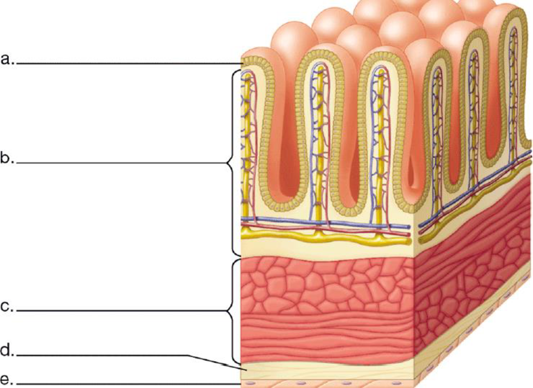
To label: The tissue layers as shown in the section of small intestine. Also to describe the concept that ‘the structure correlates with functions’; ‘an animal’s body has a hierarchy of organization with emergent properties at each level’; and ‘complex bodies have structural adaptations that increase surface area for exchange’ in the diagram.
Introduction: The structure of each specialized cell type fits to their functions. A tissue derives its function by the cells that make it up. For example,the columnar epethelial cells.
Connective tissues have fibres and cells that provide support and connection to the tissue. Therefore, the connection of cell to tissue to the organ is visible in the given diagram.
There are many projections that line the surface of the small intestine for the absorption of the nutrients. The villi increases the surface area of the small intetine for absorption of nutrients.
Answer to Problem 1CC
Pictorial representation: A labeled diagram showing different tissue layers in a section of small intestine is presented in the Fig.1.
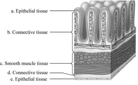
Fig.1: Different section of small intestine.
Explanation of Solution
(a)
Correct answer: Epithelial tissue.
Explanation: In the small intestine, epithelial tissue covers the outside of the villi. Hence the correct answer is epithelial tissue.
(b)
Correct answer: Connective tissue.
Explanation: Connective tissue attaches and provides support to the organs. Hence the correct answer is connective tissue
(c)
Correct answer: Smooth muscle tissue.
Explanation: Small intestine requires strength to push the digested product down its length, and this is provided by smooth muscle tissue. Hence the correct answer is smooth muscle tissue.
(d)
Correct answer: Connective tissue.
Explanation: Connective tissue attaches and provides support to the organs. Hence the correct answer is connective tissue.
(e)
Correct answer: Epithelial tissue.
Explanation: It attaches to the smooth muscle tissue and provide strength to the small intestine. Hence the correct answer is epithelial tissue.
Want to see more full solutions like this?
Chapter 20 Solutions
CAMPBEL BIOLOGY:CONCEPTS & CONNECTIONS


- Draw a plan diagram of the frog intestine viewed under the microscope (see attached images for help!!).... YOU NEED TO DO A DRAWING AND DO CALCULATIONS. DO NOT ANSWER THIS WITH A WRITTEN RESPOSE!!!! A plan diagram is a simple drawing showing only the boundaries between the lumen, the columnar epithelium, the (areolar) connective tissue, the circular smooth muscle and the longitudinal smooth muscle. A plan diagram serves to illustrate the location and relative thickness of the various tissue layers. 1. Draw a plan diagram and label the following: • Longitudinal smooth muscle • Columnar epithelial tissue • Circular smooth muscle • Villi • Areolar connective tissue • Lumen 2. Calculate actual size and drawing magnification of the width of the intestine. Include formulae used and calculations. 3. Add a scale bar with actual size next to your diagram. Give your drawing a descriptive title and record total magnification. Attached are an example plan diagram labelled, as well as a…arrow_forwardA hypothetical organ has the following functional requirements : (1) the ability to resist surface abrasion and mechanical stresses;(2) the ability to contract involuntarily when stimulated by cells of the nervous system; and (3) the ability to resist tension in many different planes of force. The organ needs one tissue to carry out each of these requirements, and it also needs one tissue to " glue" all other tissues together, and one tissue to stimulate the contracting cells. What are the five tissues that will make up this hypothetical organ ? Justify your choices .arrow_forwardCreate a concept map,(not a drawing) that connects the three systems (digestive, circulatory, and respiratory system). You need to describe what is happening in your body when you eat and breathe. ADD PICTURES for the concept map start with a picture of the mouth, and make sure to add alot of details make the concept map as informative as possible. ALSO START WITH THE MOUTH IN THE MIDDLE OF THE CONCEPT MAP Example of what a concept map looks like below:arrow_forward
- Role of Homeostasis in Human Physiology: A Review After reading the article, research what disorders result from loss of homeostasis. Choose 2 disorders, provide an overview of the diseases, characteristics of the normal homeostatic mechanisms, what aspects of the mechanisms are disrupted, and what treatments are used to restore normal homeostasis or treat the symptoms of the disorder.arrow_forwardI. Give the derivatives of the following structures (one each only): 1. foregut - ____________________ 2. neural tube - _________________ 3. pharyngeal pouch I - ___________ 4. sclerotome - __________________ 5. laryngotracheal groove - ________ 6. telencephalon - _______________ 7. hepatic diverticulum - __________ 8. metencephalon - ______________ 9. cardiac neural crest - __________ 10. myotome - __________________ 11. lateral nasal - _______________ 12. hindgut - ___________________ 13. midgut - ___________________ 14. apaxial mesoderm - __________ 15. syndetome - ________________ 16. diencephalon - ______________ 17. dermatome - ________________ 18. optic vesicles - ______________ 19. meningotome - ______________ 20. pharyngeal pouch II - ________arrow_forwardWhats would the purpose of a research study of "Consumption of whole eggs promotes greater stimulation ofpost exercise muscle protein rather than consumption of egg whites" be. How would this research study be helpful for people.arrow_forward
- Hello, please answer the following attached Biology question correctly and fully. Please make sure to "Explain your answer with respect to differences in the overall surface area to volume ratio between the two animals", as well. Thank you.arrow_forwardDraw a plan diagram of the frog intestine viewed under the microscope (see attached images for help!!).... A plan diagram is a simple drawing showing only the boundaries between the lumen, the columnar epithelium, the (areolar) connective tissue, the circular smooth muscle and the longitudinal smooth muscle. A plan diagram serves to illustrate the location and relative thickness of the various tissue layers. 1. Draw a plan diagram and label the following: • Longitudinal smooth muscle • Columnar epithelial tissue • Circular smooth muscle • Villi • Areolar connective tissue • Lumen 2. Calculate actual size and drawing magnification of the width of the intestine. Include formulae used and calculations. 3. Add a scale bar with actual size next to your diagram. Give your drawing a descriptive title and record total magnification. Attached are an example plan diagram labelled, as well as a microscopic image of the frog intestine (3-4 cells should fit across only) PLEASE DO THE DRAWING AND…arrow_forwardDiscuss the organization of animal bodies from the cellular to the organsystem level. Given that organ systems interact extensively with eachother, should each system really be considered distinct, or is it possibleto think of an animal’s body as being composed of one large, integrated“system”?arrow_forward
- Compare the following animals in terms of their digestive tracts (centerpoint only needed in the graph (similarities))arrow_forwardCreate a concept map, that connects the three systems (digestive, circulatory, and respiratory system). You need to describe what is happening in your body when you eat and breathe. ADD PICTURES and for the concept map start with a picture of the mouth, and make sure to add alot of details make the concept map as informative as possible.arrow_forward2. List 5 subspecialties of anatomy and physiology and describe each. 3. Enumerate and discuss the six structural and functional organizations of the human body. 4. List and describe the four tissue types. Give examples. 5. In a table form, classify the organs and the organ system. Relate each of their function. 6. Distinguish between metabolism, anabolism, and catabolism. 7. Provide at least two examples of human responsiveness and human movement. 8. Compare and contrast growth, differentiation, and reproduction. 9. Describe the negative and positive feedback mechanisms. Give an example for each. 10. Using a diagram, discuss the three components of a negative feedback mechanism to maintain homeostasis.arrow_forward
 Biology: The Dynamic Science (MindTap Course List)BiologyISBN:9781305389892Author:Peter J. Russell, Paul E. Hertz, Beverly McMillanPublisher:Cengage Learning