Videos
HeLa Cells Are a Genetic Mess HeLa cells can vary in chromosome number. Defects in proteins that orchestrate cell division result in descendant cells with too many or too few chromosomes, an outcome that is one of the ha1lmarks of cancer cells. The panel of chromosomes in FIGURE 11.9, originally published in 1989, shows all of the chromosomes in a single metaphase HeLa cell.
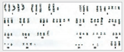
FIGURE 11.9 Karyotype of HeLa showing chromosomes in one cell.
Can you tell that this cell came from a female? How?

Trending nowThis is a popular solution!

Chapter 11 Solutions
Biology: The Unity and Diversity of Life (MindTap Course List)
Additional Science Textbook Solutions
Anatomy & Physiology
Microbiology: Principles and Explorations
Campbell Biology: Concepts & Connections (9th Edition)
Anatomy & Physiology: The Unity of Form and Function
Essentials of Human Anatomy & Physiology (11th Edition)
Becker's World of the Cell (9th Edition)


- HeLa Cells Are a Genetic Mess HeLa cells can vary in chromosome number. Defects in proteins that orchestrate cell division result in descendant cells with too many or too few chromosomes, an outcome that is one of the ha1lmarks of cancer cells. The panel of chromosomes in FIGURE 11.9, originally published in 1989, shows all of the chromosomes in a single metaphase HeLa cell. FIGURE 11.9 Karyotype of HeLa showing chromosomes in one cell. How many extra chromosomes does this cell have, compared to a normal human body cell?arrow_forwardFigure 10.6 Which of the following is the correct order of events in mitosis? Sister chromatids line up at the metaphase plate. The kinetochore becomes attached to the mitotic spindle. The nucleus reforms and the cell divides. Cohesin proteins break down and the sister chromatids separate. The kinetochore becomes attached to the mitotic spindle. Cohesin proteins break down and the sister chromatids separate. Sister chromatids line up at the metaphase plate. The nucleus reforms and the cell divides. The kinetochore becomes attached to the cohesin proteins. Sister chromatids line up at the metaphase plate. The kinetochore breaks down and the sister chromatids separate. The nucleus reforms and the cell divides. The kinetochore becomes attached to the mitotic spindle. Sister chromatids line up at the metaphase plate. Cohesin proteins break down and the sister chromatids separate. The nucleus reforms and the cell divides.arrow_forwardHeLa Cells Are a Genetic Mess HeLa cells can vary in chromosome number. Defects in proteins that orchestrate cell division result in descendant cells with too many or too few chromosomes, an outcome that is one of the ha1lmarks of cancer cells. The panel of chromosomes in FIGURE 11.9, originally published in 1989, shows all of the chromosomes in a single metaphase HeLa cell. FIGURE 11.9 Karyotype of HeLa showing chromosomes in one cell. What is the chromosome number of this HeLa cell?arrow_forward
- Figure 6.4 Which of the following is the correct order of events in mitosis? a. Sister chromatids line up at the metaphase plate. The kinetochore becomes attached to the initotic spindle. The nucleus re-forms and the cell divides. The sister chromatids separate. b. The kinetochore becomes attached to the mitotic spindle. The sister chromatids separate. Sister chromatids line up at the metaphase plate. The nucleus re-forms and the cell divides. c. The kinetochore becomes attached to metaphase plate. Sister chromatids line up at the metaphase plate. The kinetochore breaks down and the sister chromatids separate. The nucleus re-forms and the cell divides. d. The kinetochore becomes attached to the mitotic spindle. Sister chromatids line up at the metaphase plate. The kinetochore breaks apart and the sister chromatids separate. The nucleus re-forms and the cell divides.arrow_forwardA diploid organism has chromosomes. Use colours or symbols to show which chromosomes are which throughout each stage. Draw and label the chromosomes, sister chromatids, centromeres, spindle fibres, tetrad, crossing over and nucleus where appropriate. Prophase I Metaphase I Anaphase I Telophase I Prophase II Metaphase II Anaphase II Telophase II O曲T回A@ 28 Ai MacBook Pro K 2$ & 2 3 E R Y D F G K V B M command MOSISO commandarrow_forwardThe figure below depicts cells from the same organism. Cell A is demonstrating which of the following: K J A Anaphase I Anaphase II O Anaphase of mitosis O Non-disjunction LLL B دے Carrow_forward
- In dolphins, 2n=44. Drag each term or phrase to the correct position. (Each box is used once.) Somatic cell in anaphase of mitosis Ovum Somatic cell during interphase Zygote Sperm Cells with 22 chromosomes Cells with 44 chromosomes Cells with 88 chromosomesarrow_forwardThe figure below shows the number of chromosomes observed in an actively dividing cell at each stage of cell division. A B number of chromsomes per cell C 100 90 D 80 A bar graph comparing the number of chromosomes at different stages of cell division. 20 Which of the following best explains the change in the number of chromosomes between metaphase and anaphase? prophase metaphase anaphase telophase cytokinesis stage of cell division New chromosomes formed during prophase are doubled during anaphase. DNA replication occurs between metaphase and anaphase, doubling the number of chromosomes. During metaphase, a cell contains identical copies of each chromosome, and then trans- forms into sister chromatids. During anaphase, the chromatids are separated, each becoming independent chromo- somes in its respective new cellarrow_forwardStructure P Cell S State the chromosomal number in parent cell and the daughter cells formed. Parent cell: Daughter cell 1: Daughter cell 2:arrow_forward
- Mitosis is used to make more body cells while meiosis is only used to make gametes for sexual reproduction. Use the diagram to identify one similarity and one difference between mitosis and meiosis. Your answer must specifically refer to this diagram. [i.e. say what specific cells in the diagram show the similarity or difference] Mitosis Parent cell Meiosis Parent cell DNA replicates DNA replicates 2 daughter cells 2 daughter cells 4 daughter cells U.S. National Library of Medicine (Level 3) tv МacВook Air 80 DD F5 F7 F8 F9 F10 F2 F3 @ # $ & * 3 4 9. 7 8 W E R Y 一 S H J K C V M Varrow_forwardmitosis nuclei resides in the nucleus karyotype autosomes haploid In dividing parent cell; in the parent cell. Male and female gametes at fertilization forms a homologs Microscopic observations showed that during fertilization, the unite, providing evidence that genetic material resides in the cell membrane Brightly staining, threadlike bodies within the nucleus were termed of these bodies could be traced through cell division. chromosomes diploid A image can reveal abnormalities in meiosis of eggs and sperm ; the movements gametes contain a single set of chromosomes. The fusion of zygote. the number of chromosomes in daughter cells remains the same as in the the number of chromosomes in daughter cells is half that of Two chromosomes that match in size, shape, and banding when isolated and stained are is an image of an individual's chromosomes arranged in homologous pairs. This chromosome number and structure nondisjunctionarrow_forwardAssume that cell does mitosis Assume that cell does meiosis What is the diploid number of chromosomes in this organism? Give the number of chromosomes and the number of DNA molecules per cell present at the stages represented. I tried to solve them, but I'm not sure what I did right and what was wrong. Please correct it for me. See that attached photo.arrow_forward
 Biology: The Unity and Diversity of Life (MindTap...BiologyISBN:9781337408332Author:Cecie Starr, Ralph Taggart, Christine Evers, Lisa StarrPublisher:Cengage Learning
 Biology: The Unity and Diversity of Life (MindTap...BiologyISBN:9781305073951Author:Cecie Starr, Ralph Taggart, Christine Evers, Lisa StarrPublisher:Cengage Learning
 Biology Today and Tomorrow without Physiology (Mi...BiologyISBN:9781305117396Author:Cecie Starr, Christine Evers, Lisa StarrPublisher:Cengage Learning
 Biology 2eBiologyISBN:9781947172517Author:Matthew Douglas, Jung Choi, Mary Ann ClarkPublisher:OpenStax
 Concepts of BiologyBiologyISBN:9781938168116Author:Samantha Fowler, Rebecca Roush, James WisePublisher:OpenStax College