An Illustrated Guide To Vet Med Term
4th Edition
ISBN: 9781305465763
Author: ROMICH
Publisher: Cengage
expand_more
expand_more
format_list_bulleted
Question

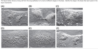
Transcribed Image Text:The figure below shows a living cell from the lung epithelium of a newt at different stages in M phase. Identify the stage in M phase that each panel in the
figure represents.
(A)
(B)
(C)
(D)
(E)
(F)
Expert Solution
This question has been solved!
Explore an expertly crafted, step-by-step solution for a thorough understanding of key concepts.
This is a popular solution
Trending nowThis is a popular solution!
Step by stepSolved in 2 steps

Knowledge Booster
Learn more about
Need a deep-dive on the concept behind this application? Look no further. Learn more about this topic, biology and related others by exploring similar questions and additional content below.Similar questions
- Give the following (a.) cell, (b.) description, and (c.) type of fiber present in tissues below: 1. Mesenchyme 2. Wharton's jelly of umbilical cordarrow_forwardDuring a microscopy exercise in the anatomy laboratory,a student makes the following observations about a tissuesection: (1) The section contains some different types ofscattered protein fibers—that is, they exhibit differentwidths, some are branched, some are long and unbranched,and their staining characteristics differ (some are seenonly with specific stains). (2) Several cell types withdifferent morphologies are scattered throughout the section,but these cells are not grouped tightly together. (3) Theexamined section has some “open spaces”—that is, placesbetween cells and the observed fibers in the section thatappear clear with no recognizable features. What type oftissue is the student observing? Where might this tissue befound in the body?arrow_forwardwhich of the following would be false? a) summation of B and C would not change membrane b) summation of B would be an IPSP c) summation of C and A = suprathreshold stimuli d) stimulation by A would depolarize cell e) repeated stimulation by A could spatially summate and reach threshold (This would be temporal summation)arrow_forward
- If you are given a prepared slide of fish gill tissue (gills are responsible for gas exchange between the fish and the surrounding water), would you expect to find simple squamous epithelium, stratified squamous epithelium, or simple columnar epithelium in the sample? (Choose one.) Explain your reasoningarrow_forwardIdentify A (name of the layer; blank 1) - Identify B (name of the structure; blank 2) - Identify C (name of the layer; blank 3) - Identify D (name of the cells; blank 4) - E: identify this organ. High mag. Low high mag. D. B B E: Identify this organ Blank # 1 Blank # 2 Blank # 3 Blank # 4 Blank # 5arrow_forwardIn the lungs of smokers, a process called metaplasia occurs in which the normal lining cells of the lung are replaced by squamous metaplastic cells (many layers of squamous epithelial cells). Functionally, why is this an undesirable body reaction to tobacco smoke? HINT Your answer should mention the structure and function of the normal lining cells of the lung.arrow_forward
- Why is the pseudostratified epithelium of the respiratory tract ciliated while the same type of tissue in the digestive tract is not ciliated?arrow_forwardA hypothetical organ has the following functional requirements : (1) the ability to resist surface abrasion and mechanical stresses;(2) the ability to contract involuntarily when stimulated by cells of the nervous system; and (3) the ability to resist tension in many different planes of force. The organ needs one tissue to carry out each of these requirements, and it also needs one tissue to " glue" all other tissues together, and one tissue to stimulate the contracting cells. What are the five tissues that will make up this hypothetical organ ? Justify your choices .arrow_forwardFollowing is a list of tissues that have specialized functions and demonstrate corresponding specialization of subcellular structure. Match the tissue with the letter of the cell structures and organelles listed to the right that would be abundant in these cells. Tissues -Enzyme (protein)-secreting cells of the pancreas -Insect flight muscles -Cells lining the respiatory passages -White blood cells that engulf and destroy invading bacteria -Leaf cells of cacti Cell Structures and Organelles: a. plasma membrane b. mitochondria c. Golgi apparatus d. chloroplast e. endoplasmic reticulum f. cilia and flagella g. vacuole h. ribosome i. lysosomearrow_forward
- A 50-year-old man comes to the physician because of a cough productive of large quantities of mucus for 6 months. He has smoked 1 pack of cigarettes daily for 25 years. Which of the following cell types is the most likely cause of the increase in this patient's secretion of mucus? (A) Columnar ciliated epithelial cells (B) Goblet cells (C) Interstitial cells (D) Macrophages (E) Pneumocyte epithelial cellsarrow_forwardRETICULAR CONNECTIVE TISSUE Describe this tissue. A. ["contains collagen fibers arranged "regularly" in bundles", B."fine interlacing network of reticular fibers and reticular cells", C."fine interlacing network of elastic fibers and chondrocytes"] What is the location where this tissue can be found? A. ["stroma of liver, spleen, lymph nodes; red bone marrow; reticular lamina of basement membrane; around blood vessels and muscles", B."forms tendons, most ligaments, aponeuroses", C."fasciae, reticular region of the dermis, pericardium, periosteum, perichondrium, joint capsules, membrane capsules around organs, heart valves"] What is the function of this tissue type? A. ["strength, elasticity, support", B."tensile strength", C."forms stroma of organs; binds smooth muscle tissue; filters and removes work out blood cells in spleen and microbes in lymph nodes"] ADIPOSE CONNECTIVE TISSUE What is the main cell type in this tissue?…arrow_forwardName the cell body of nerve cell.arrow_forward
arrow_back_ios
SEE MORE QUESTIONS
arrow_forward_ios
Recommended textbooks for you
