
Laboratory Manual For Human Anatomy & Physiology
4th Edition
ISBN: 9781260159363
Author: Martin, Terry R., Prentice-craver, Cynthia
Publisher: McGraw-Hill Publishing Co.
expand_more
expand_more
format_list_bulleted
Concept explainers
Videos
Textbook Question
Chapter 64, Problem F64.9A
Observe the human torso model and figures 64.2b, 6.4.9, and 64.10 of a cadaver. Locate the labeled features and identify the numbered features that were also identified in the fetal pig dissection.
FIGURE 64.9 Identify the arteries and veins indicated on this anterior view of the abdomen of a cadaver, using the terms provided.
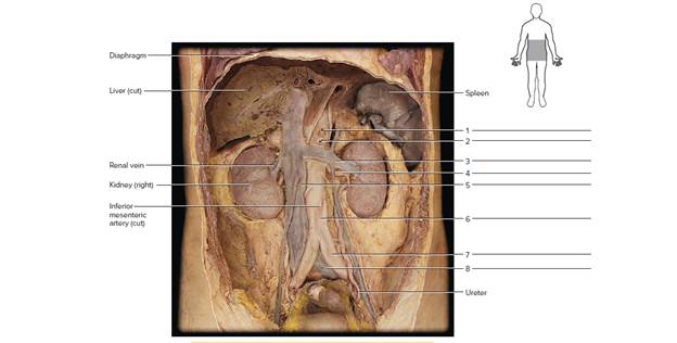
Terms:
Abdominal aorta Inferior vena cava
Celiac trunk (artery) Renal artery
Common iliac artery Renal vein
Common iliac vein Superior mesenteric artery
Expert Solution & Answer
Want to see the full answer?
Check out a sample textbook solution
Students have asked these similar questions
complete the illustration by correctly placing the following items.
abdominal acromial anterbrachial, axillary
Answer the following:
After applying the tourniquet on the antecubital fossa of a 30-year-old female patient, you were puzzled. There was no sign of the median cubital vein. You then moved to the cephalic vein, selecting a site and cleaning it. After it dried, you inserted the needle using an evacuated tube system. Blood dripped very slowly into the tube; at this rate, you thought the collection would take over 5 minutes, and the sample would likely still be inadequate. You explained this to your patient, removed the needle, and inserted it further up the vein—still almost no flow. You then reapplied the tourniquet, chose a site in the basilic vein and tried again. This time there was no blood—you must have missed the vein. What did you do wrong? What should you do now?
Answer in 10 sentences of what did you do wrong?
Answer in 10 sentences of what should you do now?
Answer the following:
After applying the tourniquet on the antecubital fossa of a 30-year-old female patient, you were puzzled. There was no sign of the median cubital vein. You then moved to the cephalic vein, selecting a site and cleaning it. After it dried, you inserted the needle using an evacuated tube system. Blood dripped very slowly into the tube; at this rate, you thought the collection would take over 5 minutes, and the sample would likely still be inadequate. You explained this to your patient, removed the needle, and inserted it further up the vein—still almost no flow. You then reapplied the tourniquet, chose a site in the basilic vein and tried again. This time there was no blood—you must have missed the vein. What did you do wrong? What should you do now?
Chapter 64 Solutions
Laboratory Manual For Human Anatomy & Physiology
Ch. 64 - Describe the relative thicknesses of the walls of...Ch. 64 - Prob. 1.2ACh. 64 - Compare the origins of the common carotid arteries...Ch. 64 - Compare the origins of the external and internal...Ch. 64 - Compare the relative sizes of the external and...Ch. 64 - Prob. 2.2ACh. 64 - Identity the numbered cardiovascular features of...Ch. 64 - FIGURE 64.8 Identify the veins on the ventral...Ch. 64 - Explain the oxygen-rich blood in the umbilical...Ch. 64 - Observe the human torso model and figures 64.2b,...
Knowledge Booster


Learn more about
Need a deep-dive on the concept behind this application? Look no further. Learn more about this topic, biology and related others by exploring similar questions and additional content below.Similar questions
- This is a cadaveric image of the thorax. Please label “E”. Look carefully at where the arrow is pointing (Label the tip of the arrow). Hint: it is not internal thoracic artery.arrow_forwardAn abnormolly is any deviation from what is regarded as normal.arrow_forwardMrs. Tillson underwent _________________ to remove excess fluid from her abdomen.arrow_forward
- When imaging the abdominal aorta in its long axis and the vena caval entry into the RA, the probe is ___________ slightly ________________.arrow_forwardChest radiographs can be obtained from two different directions (called projections); antero-posterior (AP) or posteroanterior (PA) projections. In the radiograph provided, which type of projection has been used? Explain briefly what features of the radiograph led to your decision?arrow_forwardObserve the table in the image. Use the information provided to complete the table (Anesthesia Surgery Record). If any information does not appear in the information provided, please make it up. 1- They will use the information from the attached case https://www.clinvetpeqanim.com/index.php?pag=articulo&art=230 2- HR data = 100 -110-120-100-127-122-127-127-120-110-120-127. 3- RR data = 14-14-12-8-12-14-18-18-18-12-14-18. 4- temperature data = 100-101-99-99. 5-Choose the start and end time of anesthesia 6- Set the start and end time of the surgery. 7- Choose the time of intubation and extubation. 8- Establish the oxygenation data must be between 97% 98-99-100-100-96-97-97-99-95-100-100% in a period of one hour. 9- EtCO2 = 35-38-30-22-40-32-35-38-45-45-40-45 10- sitstolica:97, 117, 110, 121, 120, 120, 106, 120, 132, 154, 120, 110 11-diastolic: 69, 77, 50, 75, 70, 80, 71, 80, 87, 93, 80, 80 12-MAP:79-97-89-89 -87 -92-87-93-96- 96-92-87arrow_forward
- During an appendectomy performed at McBurney's point, which of the following structures is most likely to be injured? * Deep circumflex femoral artery Inferior epigastric artery Illiohypogastric nerve Genitofemoral nerve Spermatic cordarrow_forwardYou are reading a surgeon’s operative report. During the course of surgery, she made several incisions. Your job is to read her operative report and determine where the incisions were made. Draw and label the incision on the figure below. The first incision was in the right anterior sternal region, 3 centimeters inferior to the cervical region. The cut extended vertically in an inferior direction, ending 2 centimeters superior to the umbilical region. The second incision began in the left anterior, lateral inguinal region and extended horizontally in a medial direction for 4 centimeters to the pubic region. At the pubic region, the cut turned and extended vertically in a distal direction for 4 centimeters and ended in the femoral region. The third incision was made in the left posterior, lateral femoral region, 2 centimeters proximal to the popliteal region. The cut extended in a distal direction to 2 centimeters proximal to the tarsal region.arrow_forwardInstructions: 1. Label the dorsal and ventral external features of the frog's heart. Answer all labels on the space provided. 1. pulmonary vein 2. 3. 4. 6 5. 6. 7. Figure 11.3 Dorsal view of the frog's heartarrow_forward
- submit a blank (unfilled) necropsy report layout for animalsarrow_forwardThe patient was given IV (intravenous) with medication into the left great saphenous vein. Describe flow of the medication through the blood vessels into the right atrium. Specify all blood vessels on the way from the left great saphenous vein into the right atrium.arrow_forwardClearly state each step you would use in preparing the injections and state the steps in the proper order. Select the correct syringe to the right and mark the dose of Humilin R and Humilin N on the syringe.arrow_forward
arrow_back_ios
SEE MORE QUESTIONS
arrow_forward_ios
Recommended textbooks for you

 Medical Terminology for Health Professions, Spira...Health & NutritionISBN:9781305634350Author:Ann Ehrlich, Carol L. Schroeder, Laura Ehrlich, Katrina A. SchroederPublisher:Cengage Learning
Medical Terminology for Health Professions, Spira...
Health & Nutrition
ISBN:9781305634350
Author:Ann Ehrlich, Carol L. Schroeder, Laura Ehrlich, Katrina A. Schroeder
Publisher:Cengage Learning
Dissection Basics | Types and Tools; Author: BlueLink: University of Michigan Anatomy;https://www.youtube.com/watch?v=-_B17pTmzto;License: Standard youtube license