
Videos
21-Year-Old Female with Skeletal Injuries
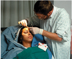
While riding her bike to campus, 21-year-old Liliana Rose was struck by a car. Examination in the Emergency Department reveals several injuries. Relative to her integumentary system, the following comments are noted on her chart:
•Epidermal abrasions of the right lateral upper arm and anterior shoulder.
•A deep, 2-cm laceration extending vertically on right lateral cheek and a horizontal 1-cm laceration on the temple.
•Cyanosis is apparent in her nail beds and lips.
The lacerated areas are cleaned, sutured (stitched), and bandaged by the emergency room (ER) personnel.
3. Liliana is concerned about scarring to her face. The surgeon tells her that the cut extending along her temple should heal with little scarring, but the one extending up her cheek is more likely to scar. Why might this be the case?
Want to see the full answer?
Check out a sample textbook solution
Chapter 5 Solutions
HUMAN A&P LL W/MOD.MAST.TCC ACCESS >IB<


- A 25-year-old man presents with complaints of scaling skin lesions over the elbow, as shown in the image attached, that are waxing and waning over the past year. On physical examination, similar lesions are present on the buttock region, trunk, and knees along with yellow discoloration and pitting of the nails. Which of the following microscopic findings is most likely seen in the skin lesions? Answers A-E A Hypergranulosis and interface dermatitis B Hyperkeratosis and keratin horn cysts C D Parakeratosis and Munro microabscess Subepidermal separation of the basal cells E Suprabasal acantholysis and fluid accumulation O O Question # 38 attachmentarrow_forwardPlease define and describe the cause of the skin abnormality: Birthmarks Albinism Vitiligo Freckles Melasma Age spots Lentigo Pellagraarrow_forwardMr. Jordan puts his call light on to request assistance to ambulate. You walk in and observe from the loose gown that he has subclavicular retractions and cyanosis of his nail beds. What would be the most appropriate nursing actions at this time? and why? PreviousNextarrow_forward
- Intradermal melanocytic pigmented nevus H&E staining Mark the corresponding elements in the picture: 1 - epidermis 2 - dermis 3- nevus-cells 4- accumulation of melanin Description: Specify the localization of melanin accumulationsarrow_forwardThis is an image of skin. Identify "5" dermis epidermis hypodermis 5arrow_forward(10) medical terms and define them in your own word. sebaceous glands sebum Perspiration Hair follicles arrector pili dermatologist plastic surgeon Acne vulgaris comedo Seborrheic dermatitisarrow_forward
- Subcutaneous Tissue or Hypodermis 1. The illustration below depicts a magnified view of the hypodermis. Label the numbered regions and structures on this illustration. 1- 2- bo' Integument Exercises 10 lo girle la si des -3 neno -4arrow_forwardSkin Disorders" ( minimum 3 disorders) and "Skin Cancers" ( minimum 3 types of Skin cancers)- History, Causes, Types, suggested treatment options and remedies asaparrow_forwardLabel the following:arrow_forward
 Essentials of Pharmacology for Health ProfessionsNursingISBN:9781305441620Author:WOODROWPublisher:Cengage
- Understanding Health Insurance: A Guide to Billin...Health & NutritionISBN:9781337679480Author:GREENPublisher:Cengage
 Medical Terminology for Health Professions, Spira...Health & NutritionISBN:9781305634350Author:Ann Ehrlich, Carol L. Schroeder, Laura Ehrlich, Katrina A. SchroederPublisher:Cengage Learning