Videos
Using clay, demonstrate how cocci can divide in several planes and show the outcome of this division. Show how the arrangements of bacilli occur, including palisades.
Introduction:
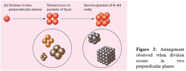
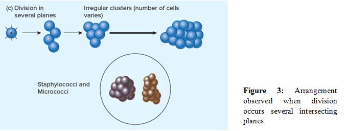
Cocci show a variety of arrangement and can also be categorized on that basis. It is mainly influenced by the pattern of division and how cells remain attached even after that.
Explanation of Solution
Bacterial cells can be classified on the basis of their arrangement, or grouping pattern. A wide variety of arrangements is seen in cocci. This is influenced by the division pattern and the attachment properties of the cell. The cells can appear as single, in pairs (diplococci), in tetrads (groups of four), irregular clusters (staphylococci and micrococci), in chains (streptococci), or in complex cubical packets (sarcina). These arrangements when coccus is divided along single plane, perpendicular planes, or in several intersecting planes.

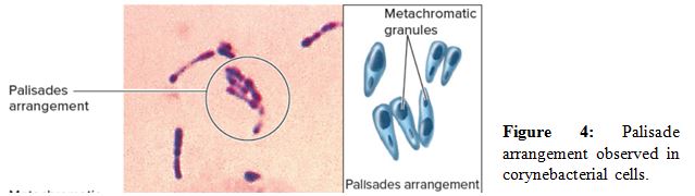
Bacilli do not exhibit wide variety in arrangement as they divide along the transverse plane. So they can occur as single cell or in pairs with their ends attached (diplobacilli), or as chains of several cells (streptobacilli). Palisade arrangement is displayed only by corynebacterial, eherre the cells remain partially attached by a small hinge region at the end. These cells tend to snap back over each other too form rows of cells arranged side by side.
Hence, on the basis of division pattern, different types of cocci arrangements are obtained and this is used as a criterion to categorize bacterial cells.
Want to see more full solutions like this?
Chapter 4 Solutions
Foundations in Microbiology


- Explain the steps for the reproduction of slime mold according to the image !arrow_forwardExplain the Arrangement of cocci resulting from different planes of cell division.arrow_forwardPlease hand draw Paramecium caudatum and label it with the following terms: Paramecium, Phylum Ciliophora, Macronucleus, micronucleus. Please be sure to include all of the terms provided.arrow_forward
- Differentiate the locations of flagella by providing illustrationsarrow_forwardDescribe the mycobacterial cell wall and give one reason why it is important in the treatment of the pathogen Mycobacterium tuberculosis. Thank youarrow_forwardBelow is an image of bacterial cells. 1. What shape of bacterial cells is portrayed? 2. Are there any bacterial arrangements visible? If so, which one(s)?arrow_forward
- Draw a labeled diagram of structure of spore of Riccia.arrow_forwardDefine arrectores pilorumarrow_forwardAnswer the following questions: 1. Is the cell flexible? 2. How can you determine the anterior and posterior end of ciliate mophologically? 3. How is the oral groove strategically advantageous for the Paramecium sp.?arrow_forward
- Labe A. CHLOROPHYTA (Green Algae) Observe the structure of algae provided in the laboratory. Label the following diagrams. (a) Chlorella (b) Chlamydomonas Differentiate the structure between genus (a) and (b).arrow_forwardWhy does whisking egg white produce foam?arrow_forwardB. PHAEOPHYTA (Brown Algae) C. RHOI Identify and label the structures of Padina and Sargassum. Padina vickersiae Padina Sem Sargassum Differentiate the structure between Padina and Sargassum.arrow_forward
