Concept explainers
Videos
To label: The lymphatic structures.
Introduction: Lymphatic system contains a group of tissues and organs which protect our body from toxins and unwanted materials. It contains well-defined structures present in our body. The primary function of the lymphatic system is to transport a lymphatic fluid containing white blood cells that fight against unwanted materials throughout the body. The red bone marrow, thymus, lymph nodes, tonsils, and spleen come under the lymphatic organs.
Answer to Problem 1.1BGL
Pictorial representation:
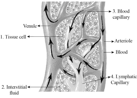
Fig 1: Lymphatic and blood capillaries
Explanation of Solution
In the above-given figure, the blood capillary, interstitial fluid, lymphatic capillary, and tissue cells are labeled.
Excess fluid in the interstitial space, space between the tissues and blood capillaries, is termed as interstitial fluid. Once, the interstitial fluid enters the lymphatic capillaries (located near the blood capillaries) then it is said to be lymph. Lymph fluid flows from the lymphatic capillaries into the lymphatic vessels.
To label: The lymphatic capillary.
Answer to Problem 1.1BGL
Pictorial representation:
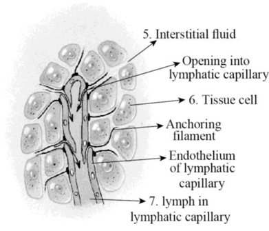
Fig 2 lymphatic capillaries
Explanation of Solution
In the above-given image, the interstitial fluid, tissue cell, and the lymph in the lymphatic capillaries are labeled.
Interstitial fluid: It is also known as tissue fluid. It is a thin layer of fluid that surrounds the body’s cells in the interstitial spaces (tissues spaces).
Lymphatic capillaries: Tiny, thin-walled vessels exist in the spaces between the cells all over the body.
To label: The lymphatic capillaries.
Answer to Problem 1.1BGL
Pictorial representation:
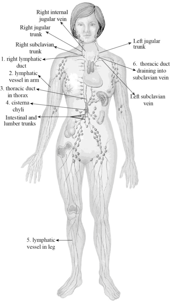
Fig 3: Lymphatic capillaries
Explanation of Solution
The fig:3 clearly indicates the structures like right lymphatic duct, lymphatic vessels in the arm, thoracic duct, cistern chili, lymphatic vessels, and thoracic duct.
Lymphatic vessels flow into lymph nodes to drain lymph. Lymph trunks join to form the thoracic duct and right lymphatic duct. The lymph trunk merges to form cisterna chyli in the abdominal region. The thoracic duct initiates at the cisterna chyli and drains the lymph from the legs, abdomen, left arm, left the side of the thorax, neck, and head.
The right lymphatic duct drains lymph from the right arm by means of the right jugular trunk, and right side of the thorax, neck, and head by means of the right subclavian trunk.
Right lymphatic duct: Also known as a right thoracic duct that drains the lymphatic fluid usually from the right thoracic cavity, right arm, neck’s right side, and the head.
Lymphatic vessels: Thin-walled vessels or tubes that carry the lymph.
Thoracic duct: It is the major vessels of the lymphatic system. It collects the large portion of the lymph and drains into the blood.
Cisterna chyli: A dilated sac located at the lower end of the thoracic duct in which the lymph flows from the interstitial trunk.
To label: The lymphatic drainage.
Answer to Problem 1.1BGL
Pictorial representation:
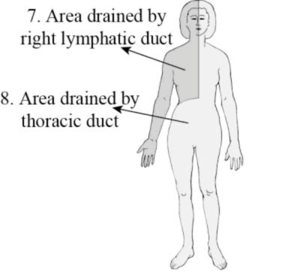
Fig 4: Lymphatic drainage
Explanation of Solution
The fig:4 clearly indicates the area drained by the right lymphatic duct and the area drained by the thoracic duct.
Lymphatic vessels flow into lymph nodes to drain lymph. Lymph trunks join to form the thoracic duct and right lymphatic duct. The lymph trunk merges to form cisterna chyli in the abdominal region. The thoracic duct initiates at the cisterna chyli and drains the lymph from the legs, abdomen, left arm, left the side of the thorax, neck, and head.
The right lymphatic duct drains lymph from the right arm by means of the right jugular trunk, and right side of the thorax, neck, and head by means of the right subclavian trunk.
To identify: The thoracic duct
Answer to Problem 1.1BGL
Pictorial representation:
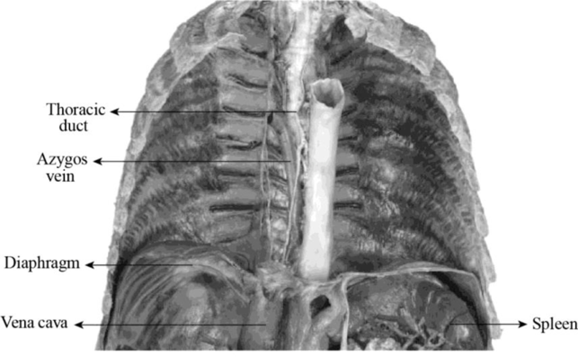
Fig: 5 Thoracic duct in cadaver
Explanation of Solution
The fig:5 clearly indicates the structures like thoracic duct, azygos vein, diaphragm, vena cava, and spleen.
Thoracic duct: It is the major vessels of the lymphatic system. It collects the large portion of the lymph and drains into the blood.
Azygos vein: It is a vein running up the side of the thoracic vertebral column. It drains itself towards the superior vena cava.
Diaphragm: A skeletal muscle located at the bottom of the chest and separates the abdomen from the chest.
Vena cava: Vein that returns the blood to the heart from various body parts.
Spleen: Largest lymphatic organ situated above the stomach in the left upper quadrant of the abdomen and under the ribcage.
Want to see more full solutions like this?
Chapter 31 Solutions
Laboratory Manual for Anatomy and Physiology, 6e Loose-Leaf Print Companion


 Human Anatomy & Physiology (11th Edition)BiologyISBN:9780134580999Author:Elaine N. Marieb, Katja N. HoehnPublisher:PEARSON
 Biology 2eBiologyISBN:9781947172517Author:Matthew Douglas, Jung Choi, Mary Ann ClarkPublisher:OpenStax
 Anatomy & PhysiologyBiologyISBN:9781259398629Author:McKinley, Michael P., O'loughlin, Valerie Dean, Bidle, Theresa StouterPublisher:Mcgraw Hill Education,
 Molecular Biology of the Cell (Sixth Edition)BiologyISBN:9780815344322Author:Bruce Alberts, Alexander D. Johnson, Julian Lewis, David Morgan, Martin Raff, Keith Roberts, Peter WalterPublisher:W. W. Norton & Company
 Laboratory Manual For Human Anatomy & PhysiologyBiologyISBN:9781260159363Author:Martin, Terry R., Prentice-craver, CynthiaPublisher:McGraw-Hill Publishing Co.
 Inquiry Into Life (16th Edition)BiologyISBN:9781260231700Author:Sylvia S. Mader, Michael WindelspechtPublisher:McGraw Hill Education