
Laboratory Manual For Human Anatomy & Physiology
4th Edition
ISBN: 9781260159363
Author: Martin, Terry R., Prentice-craver, Cynthia
Publisher: McGraw-Hill Publishing Co.
expand_more
expand_more
format_list_bulleted
Videos
Textbook Question
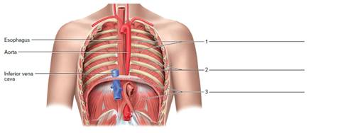
Chapter 23, Problem F23.8A
Label the muscle of respiration of the posterior body wall (anterior view). The heart and lungs are removed.
Expert Solution & Answer
Want to see the full answer?
Check out a sample textbook solution
Students have asked these similar questions
A patient whose mouth open when breathing their nasal cannula is positioned in their nose would have no oxygen delivered to their lungs.
True or False
which respiratory muscle is responsible for moving the majority of air during eupnea? sternocleidomastoid, intercostals, or diaphragm.
Label the muscles used in inspiration and forced expiration by clicking and dragging the labels to the correct location. (Some labels
may not be used.)
Rectus abdominis
Muscles of Inspiration
Muscles of Forced Expiration
Diaphragm
Scalenes
Pectoralis minor
Sternocleidomastoid
External oblique
External
Intercostals
Internal
Intercostals
Chapter 23 Solutions
Laboratory Manual For Human Anatomy & Physiology
Ch. 23 - Chest and shoulder muscles that move the arm at...Ch. 23 - Muscles located in the arm that move the forearm...Ch. 23 - Rotator cuff muscles all have origins on the a....Ch. 23 - Which of the following is not a rotator cuff...Ch. 23 - The belly of the muscle is usually ________ the...Ch. 23 - The belly of the flexor digitorumprofundus muscle...Ch. 23 - There are more muscles that move the forearm than...Ch. 23 - The action of the brachialis muscle is flexion of...Ch. 23 - FIGURE 23.7 Label the deep anterior muscles of the...Ch. 23 - Label the muscle of respiration of the posterior...
Ch. 23 - FIGURE 23.9 Identify the posterior muscles of the...Ch. 23 - FIGURE 23.10 Label the anterior muscles of the...Ch. 23 - FIGURE 23.11 Identify the anterior muscles of the...Ch. 23 - Match the muscles in column A with the actions in...Ch. 23 - Name the muscle indicated by the following...Ch. 23 - Name the muscle indicated by the following...Ch. 23 - Name the muscle indicated by the following...Ch. 23 - Name the muscle indicated by the following...Ch. 23 - Name the muscle indicated by the following...Ch. 23 - Name the muscle indicated by the following...Ch. 23 - Name the muscle indicated by the following...Ch. 23 - Name the muscle indicated by the following...Ch. 23 - Identify the muscles indicated in figure 23.12....Ch. 23 - Tendons of the rotator cuff muscles hold the head...
Knowledge Booster


Learn more about
Need a deep-dive on the concept behind this application? Look no further. Learn more about this topic, biology and related others by exploring similar questions and additional content below.Similar questions
- Which of the following muscles is closest in proximity to the tongue? digastric mylohyoid geniohyoid platysma Which rib's costal cartilage (sternocostal joint) articulates with the sternum at the manubriosternal joint? 1st rib 2nd rib 3rd rib 4th rib What is the name of the cartilage that covers the airway upon deglutition? 6 epiglottis thyroid cricoid hyalinearrow_forwardDrag a label to name each of the indicated structures on this inferior view of the diaphragm. 12th rib psoas oesophagus left phrenic nerve costal muscle fibers W 11th rib quadratus lumborum superior vena cava right phrenic nerve vertebral muscle fibers thoracolumbar fascia iliac crest aorta inferior vena cava sternal muscle fibers xiphoid process central tendonarrow_forwardName the muscle/s involved in the following actions. a) Muscles of the chest 1. To exhale in regular quiet breathing: 2. To inhale deeply: 3. To exhale forcibly:arrow_forward
- Label sartorius, adductor longus, adductor magnus, gastrocnemius, tibialis posticus, extensor cruris, tibialis anticus and peroneus. LABEL THE PICTURE. Do not just copy paste google images and put it here!arrow_forwardSectional views of the trachea and lung.arrow_forwardMatch the statement on the left with the accurate directional term on the right Drag and drop options on the right-hand side and submit. For keyboard navigation... SHOW MORE ✓ The heart is () to the larynx. ipsilateral The biceps brachii muscle is (___) to the inferior integument. The trachea is () to the esophagus. anterior The right carpus is (__) to the right pollex. deep The left kidney is () to the stomach. posterior ||| III ||| ||| X X Xarrow_forward
- Which muscles are directly involved during forceful inspiration? (Select all that apply.) Internal intercostal muscles Sternohyoid Sternocleidomastoid Scalene O Diaphragmarrow_forwardThe trachealis is a band of (Select) muscle located on the V [ Select ) of the trachea. lateral anterior medial posteriorarrow_forwardIn the paragraph choose one word from each parenthesis set according to the sentences before and after. Under conditions where normal breathing isn’t sufficient, other muscles will assist the diaphragm in changing the volume of the thoracic cavity. A muscle that increases the volume of the thoracic cavity will cause pressure to (increase/decrease) which means that it is helping with forced (inspiration/expiration). A muscle that decreases the volume of the thoracic cavity will cause pressure to (increase/decrease) which means that it is helping with forced (inspiration/expiration).arrow_forward
- Jackie is a professional singer. Her singing tasks require her to allocate only 1 second for inhalation and longer expiration for prolonged notes while performing. Hence, there is a need to elevate intra-abdominal pressure to improve control during long utterances. All these muscles are affected in this activity. SELECT ALL THAT APPLY. * Rectus abdominis V Diaphragm Intercostal muscles External oblique Which respiratory center/s is/ are used by Jackie (from previous item) for elevating the intra- abdominal pressure?* Dorsal respiratory group Ventral respiratory group Pontine respiratory group DRG & VRG DRG, VRG, & PRGarrow_forwardDevelopment of the lungarrow_forwardAnterior view Posterior view 6. hyoid bone 7. trachealis muscle Gross structures of the larynx (structures may be shown more than once): cricoid cartilage epiglottis laryngeal prominence thyroid cartilage tracheal cartilagearrow_forward
arrow_back_ios
SEE MORE QUESTIONS
arrow_forward_ios
Recommended textbooks for you
 Fundamentals of Sectional Anatomy: An Imaging App...BiologyISBN:9781133960867Author:Denise L. LazoPublisher:Cengage Learning
 Comprehensive Medical Assisting: Administrative a...NursingISBN:9781305964792Author:Wilburta Q. Lindh, Carol D. Tamparo, Barbara M. Dahl, Julie Morris, Cindy CorreaPublisher:Cengage Learning
Fundamentals of Sectional Anatomy: An Imaging App...
Biology
ISBN:9781133960867
Author:Denise L. Lazo
Publisher:Cengage Learning
Comprehensive Medical Assisting: Administrative a...
Nursing
ISBN:9781305964792
Author:Wilburta Q. Lindh, Carol D. Tamparo, Barbara M. Dahl, Julie Morris, Cindy Correa
Publisher:Cengage Learning
The Musculoskeletal System | Educational Videos for Kids; Author: Happy Learning English;https://www.youtube.com/watch?v=ynVRDsDC-84;License: Standard youtube license