Connecting the Concepts
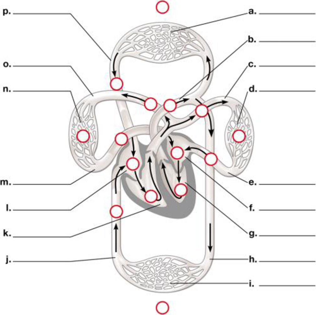
1. Use the following diagram to review the flow of blood through a human cardiovascular system. Label the indicated parts, highlight the vessels that carry oxygen-rich blood, and then trace the flow of blood by numbering the circles from 1 to 10, starting with 1 in the right ventricle. (When two locations are equivalent in the pathway, such as right and left lung capillaries or capillaries of top and lower portion of the body, assign them the same number.)

To label: The diagram of the flow of blood through the circulatory system.
Concept introduction:
The blood flows through the circulatorysystem. It includes various capillaries, aorta, pulmonary artery, pulmonary veins, ventricles, auricles, superior and inferior vena cava.
Answer to Problem 1CC
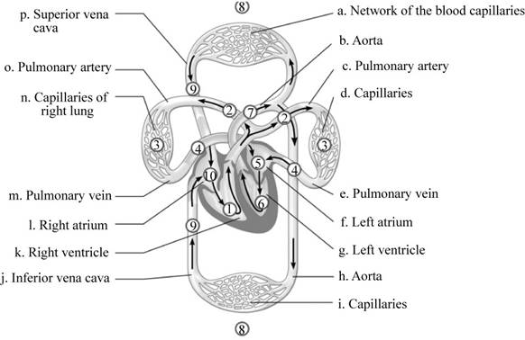
Pictorial representation: A labelled diagram of human circulatory system showing the flow of blood through it is given on Fig.1.

Fig. 1: The circulatory system.
Explanation of Solution
The labeling of the blood vessels in the circulatory system include as follows:
(a)
Network of the blood capillaries: It supplies blood to the various parts of the human body like head, arms, chest and legs.
(b)
Aorta: The blood moves from the heart chambers through this blood vessel.
(c)
Pulmonary artery: It carries the deoxygenated blood.
(d)
Capillaries: They supply blood to the lungs: The blood flows through them and reaches the lungs.
(e)
Pulmonary vein: It carries oxygenated blood.
(f)
Left atrium: It is the upper chamber of the heart which contracts to cause the flow of the blood out of the heart.
(g)
Left ventricle: It is the lower chamber of the heart where the blood flows.
(h)
Aorta: This blood vessel transports the blood to the organs from the heart.
(i)
Capillaries: They supply the blood of abdominal regions and legs:
(j)
Inferior vena cava: It drains the blood into the right atrium.
(k)
Right ventricle: It is the lower chamber of the heart.
(l)
Right atrium: It is the upper chamber of the heart.
(m)
Pulmonary vein: The left atrium of the heart receives blood from the pulmonary vein.
(n)
Capillaries of right lung: It helps in the flow of the blood to the lungs.
(o)
Pulmonary artery: It carries deoxygenated blood.
(p)
Superior vena cava: It circulates the blood in the sino-atrial node in the right atrium.
Want to see more full solutions like this?
Chapter 23 Solutions
Campbell Biology: Concepts & Connections (8th Edition)
- Figure 40.11 Which of the following statements about the heart is false? The mitral valve separates the left ventricle from the left atrium. Blood travels through the bicuspid valve to the left atrium. Both the aortic and the pulmonary valves are semilunar valves. The mitral valve is an atrioventricular valve.arrow_forwardjust answer the circled questionarrow_forward2. Make a table listing all the parts and functions of the Circulatory system. the 3. Describe the process done during Pulmonary and Systemic Circulation. Use illustration provided below. Source: Human Physiology from Cells to Systems seventh Capilary bed of lungs where gas exchange occurs Pulmonary arteries Pulmonary veins Pulmonary circuit Aorta and branches Vena cavae Left atrium Left ventricle Right atrium Right ventricle Systemic arteries Systemic veins Охудел роог, CO, - rich błood Oxygen rich, CO, - poor blood Systemic circuit Capilary bed of all body tissues where Lesson 1.: item gas exchange Occurs |Pagearrow_forward
- 5. Describe the conductions of electrical signals through the heart. answer this question with a narrative of the structures involved in initiating and coordinating the contraction of the heart. You need to include the function of each structure. Mention pacemaker cells, the sinoatrial node, atrioventricular node, internodal pathway, bundle of His, bundle branches, Purkinje fibers, first degree block, second degree block, and third degree block.arrow_forwardLet's Review - Human Circulation Your textbook begins the discussion of the path that blood takes around the body starting at the right atrium. But remember that this path a cycle, and therefore there is no true beginning or end. To make sure you have the idea, I would like you to trace the flow of blood around the human body starting at the AORTA. Complete the activity below. 1 E Right Atrium E Right ventricle 3 Aorta 4 Left Ventricle 5 : Left Atrium 6 Vena Cava 7 | Cells of the body 8 | Lungs ::::arrow_forwardPut the following in proper order of blood flow for pulmonary and systemic circulation. 1 [ Choose ] [Choose ] Capillaries at the systemic organs (e.g. liver or kidney) Left side of the heart Right side of the heart Lungs 3 [ Choose ] 4 [ Choose ]arrow_forward
- Conceptualize in Color Principal Veins Identify and color the principal veins in the figure using the colors suggested, or choose your own. • Basilic: Purple • Cephalic: Orange • Axillary: Pink • Subclavian: Blue • Brachiocephalic: Green • Superior vena cava: Dark red • Internal jugular: Orange • External jugular: Purple • Median cubital: Green • Inferior vena cava: Tan • External iliac: Red • Internal iliac: Dark blue • Common iliac: Yellow • Hepatic: Light red • Great saphenous: Brown • Posterior tibial: Orange • Anterior tibial: Gold • Popliteal: Pink • Femoral: Purple Chapter 16 Vascular System 207arrow_forwardPut these in orderarrow_forwardThe blood in the human body flows from the Blank 1. Fill in the blank, read surrounding text. atrium through Blank 2. Fill in the blank, read surrounding text. valve to left ventricle, then through semilunar valve to the Blank 3. Fill in the blank, read surrounding text. , through large and smaller arteries and arterioles to capillaries in all parts of the body, then through venules and larger and larger veins to vena Blank 4. Fill in the blank, read surrounding text. , from there to right atrium, through Blank 5. Fill in the blank, read surrounding text. valve to right ventricle, through Blank 6. Fill in the blank, read surrounding text. valve to Blank 7. Fill in the blank, read surrounding text. trunk and arteries, through the capillaries in the alveoli, to pulmonary Blank 8. Fill in the blank, read surrounding text. , then we are back at the beginning in the left atrium. Word list: left, right, mitral, semilunar, aorta, brachiocephalic, saphena, azygous, cava, tricuspid,…arrow_forward
- 2. Put the following structures in the proper order (#1-21) in which blood flows through the cardiovascular system. 1 Right atrium Left ventricle 21 Vena cava Pulmonary trunk Tricuspid valve Systemic capillaries Pulmonary capillaries 19 Systemic venules Pulmonary arteries Right ventricle Aortic valve Aorta Pulmonary veins Mitral valve Systemic veins 17 Systemic arterioles _9_ Pulmonary venules Pulmonary valve Left atrium Systemic arteries Pulmonary arterioles 3. Identify structures of the heart on the following pictures. A. В. C. D. B E. D. F. Н. G I. J. A. B. -E C. D. E. F. G. H. I. J.arrow_forwardGive a term to describe the statement. All valves are closed and the volume of blood remains constant within the relaxing ventricles.arrow_forwardII. Induction similar or relatea, the fourth does not belong with the other three. Choose the one term that does NOT belong in each of the following groupings In each question, three of the anatomical and physiological terms are A D Pulmonary trunk Right side of the Left side of the heart Vena cava 16 heart Electrical 17 T wave Pwave activity of the ventricles QRS wave AV valves Semilunar valves open AV valves Ventricular 18 closed opened systole 19 Tricuspid valve Mitral valve Bicuspid valve Left AV valve Pulmonary Superior Vena Cava 20 Umbilical Artery Pulmonary vein arteryarrow_forward
 Biology 2eBiologyISBN:9781947172517Author:Matthew Douglas, Jung Choi, Mary Ann ClarkPublisher:OpenStax
 Human Physiology: From Cells to Systems (MindTap ...BiologyISBN:9781285866932Author:Lauralee SherwoodPublisher:Cengage Learning


