Human Anatomy & Physiology (11th Edition)
11th Edition
ISBN: 9780134580999
Author: Elaine N. Marieb, Katja N. Hoehn
Publisher: PEARSON
expand_more
expand_more
format_list_bulleted
Question

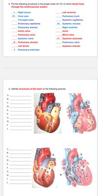
Transcribed Image Text:2. Put the following structures in the proper order (#1-21) in which blood flows
through the cardiovascular system.
1 Right atrium
Left ventricle
21
Vena cava
Pulmonary trunk
Tricuspid valve
Systemic capillaries
Pulmonary capillaries
19
Systemic venules
Pulmonary arteries
Right ventricle
Aortic valve
Aorta
Pulmonary veins
Mitral valve
Systemic veins
17
Systemic arterioles
_9_ Pulmonary venules
Pulmonary valve
Left atrium
Systemic arteries
Pulmonary arterioles
3. Identify structures of the heart on the following pictures.
A.
В.
C.
D.
B
E.
D.
F.
Н.
G
I.
J.
A.
B.
-E
C.
D.
E.
F.
G.
H.
I.
J.
Expert Solution
This question has been solved!
Explore an expertly crafted, step-by-step solution for a thorough understanding of key concepts.
This is a popular solution
Trending nowThis is a popular solution!
Step by stepSolved in 2 steps

Knowledge Booster
Similar questions
- As we auscultate the heart it is imperative, we understand what part of the heart we are listening to. Cellular senescence is observed when there is obstruction of the coronary circulation to the muscle tissue of the heart. If the left anterior descending artery (a.k.a. “Widow maker”) is occluded, predict an alternative route of blood flow to the myocardium. Start from the left coronary artery.arrow_forward#3arrow_forward3. Which heart sound is higher pitched and shorter in duration? Lub or Dup Closure of what valves (and the resulting vibrations) produce this sound?arrow_forward
- 8. We discussed fish, amphibian, and mammalian hearts, but didn’t spend much time on reptiles and birds. Please compare and contrast the anatomy of the heart ina) most reptiles (turtles, snakes, lizards) in terms of number of atria, number of ventricles, total number of chambers, and development of the aorta from aortic arches (left, right, or both). Use a table, not text. Also, include a drawing showing the basic template of dorsal aorta, ventral aorta, and six aortic arches, along with drawings showing the modified patterns of aortic arches in a-d above.arrow_forward1. You should be able to distinguish two heart sounds. The first sound is when the ventricle begins contracting (ventricular systole) the atrioventricular valves snap shut. This closing produces a low-pitched sound, the "lub" sound. The atrioventricular valves close when the ventricles contract due to pressure coming from the blood in the: Group of answer choices veins ventricles arteries atria 2. The "lub" sound is accompanied by the pulmonary and aortic (semilunar) valves opening. This, too, happens when the ventricles contract. These valves open due to pressure of the blood coming from the: Group of answer choices atria ventricles arteries veins 3. The second sound you hear in the heartbeat is when the ventricles begin to relax (diastole). The closing of the pulmonary and aortic (semilunar) valves produces a sound, the "dub" sound. These valves close when the ventricles relax due to the pressure of the blood coming from the: Group of answer choices atria…arrow_forwardHQ6. Put the labels to the location of the structure being described.arrow_forward
- 18. Under the microscope, I can see a section through one of the chambers of the heart. The myocardium is thick and the endocardium is very thin. In addition, I can see a valve that connects this chamber to the next which has two cusps that prevent backflow from one chamber into another. Where in the heart am 1? What does the thickness of the myocardium tell you about this region of the heart? What is the valve called? What does it do? What other valve would you see in this area of the heart? Where is it situated?arrow_forward#7arrow_forward10. Segment representing the time heart muscle is electrically silent 18. Waveform representing total ventricular SYSTOLE 19. Repolarization of the atria is found within which wave 20. Firing of the AV node occurs at the apex of which wave 19. Repolarization of the atria is found within which wave 20. Firing of the AV node occurs at the apex of which wavearrow_forward
- #11arrow_forward5. Describe the conductions of electrical signals through the heart. answer this question with a narrative of the structures involved in initiating and coordinating the contraction of the heart. You need to include the function of each structure. Mention pacemaker cells, the sinoatrial node, atrioventricular node, internodal pathway, bundle of His, bundle branches, Purkinje fibers, first degree block, second degree block, and third degree block.arrow_forwardComplete the pathway of blood... RIGHT ATRIUM FOOT | PULMONARY VALVE RIGHT VENTRICLE INFERIOR VENA CAVA SUPERIOR VENA CAVA MITRAL Leave PULMONARY VALVE VEINS one TRICUSPID AORTIC LEFT LEFT here i PULMONARY LUNGS VALVE VALVE VENTRICLE ATRIUM ARTERIES DRAGO,arrow_forward
arrow_back_ios
SEE MORE QUESTIONS
arrow_forward_ios
Recommended textbooks for you
 Human Anatomy & Physiology (11th Edition)Anatomy and PhysiologyISBN:9780134580999Author:Elaine N. Marieb, Katja N. HoehnPublisher:PEARSON
 Anatomy & PhysiologyAnatomy and PhysiologyISBN:9781259398629Author:McKinley, Michael P., O'loughlin, Valerie Dean, Bidle, Theresa StouterPublisher:Mcgraw Hill Education,
 Human AnatomyAnatomy and PhysiologyISBN:9780135168059Author:Marieb, Elaine Nicpon, Brady, Patricia, Mallatt, JonPublisher:Pearson Education, Inc.,
 Anatomy & Physiology: An Integrative ApproachAnatomy and PhysiologyISBN:9780078024283Author:Michael McKinley Dr., Valerie O'Loughlin, Theresa BidlePublisher:McGraw-Hill Education
 Human Anatomy & Physiology (Marieb, Human Anatomy...Anatomy and PhysiologyISBN:9780321927040Author:Elaine N. Marieb, Katja HoehnPublisher:PEARSON

Human Anatomy & Physiology (11th Edition)
Anatomy and Physiology
ISBN:9780134580999
Author:Elaine N. Marieb, Katja N. Hoehn
Publisher:PEARSON

Anatomy & Physiology
Anatomy and Physiology
ISBN:9781259398629
Author:McKinley, Michael P., O'loughlin, Valerie Dean, Bidle, Theresa Stouter
Publisher:Mcgraw Hill Education,

Human Anatomy
Anatomy and Physiology
ISBN:9780135168059
Author:Marieb, Elaine Nicpon, Brady, Patricia, Mallatt, Jon
Publisher:Pearson Education, Inc.,

Anatomy & Physiology: An Integrative Approach
Anatomy and Physiology
ISBN:9780078024283
Author:Michael McKinley Dr., Valerie O'Loughlin, Theresa Bidle
Publisher:McGraw-Hill Education

Human Anatomy & Physiology (Marieb, Human Anatomy...
Anatomy and Physiology
ISBN:9780321927040
Author:Elaine N. Marieb, Katja Hoehn
Publisher:PEARSON