Concept explainers
Videos
To match: The given anatomical terms with the organs/structures of the urinary system.
Introduction: The urinary system is a series of complex structures responsible for eliminating waste from the body, regulating blood volume and blood pressure, controlling levels of electrolytes and metabolites, and to regulate blood pH. The urinary system, the lungs, skin, and intestines are together to maintain the balance of chemicals and water in the body.
Answer to Problem 1P
Pictorial representation: Fig.1 shows a diagram of the organs/structures of the urinary system labeled with the given anatomical terms.
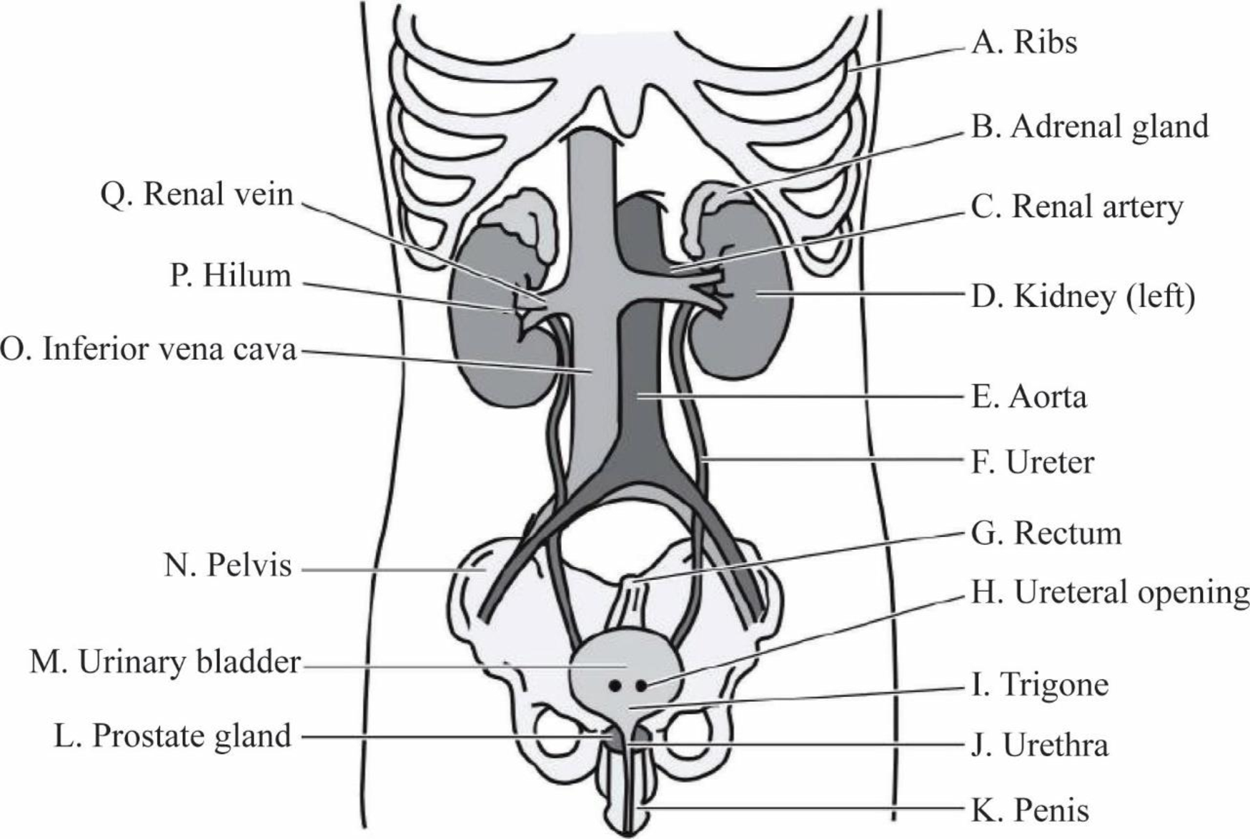
Fig 1: Anatomical structure of the urinary system
Explanation of Solution
A. Ribs: The ribs are elastic arches of bone, twelve in number on either side, separated by spaces called intercostal spaces. The main function of the ribs is to protect the kidney and liver from any external damage.
B. Adrenal gland: The adrenal glands are tiny organs that rest on top of each kidney. The right adrenal gland is pyramid shaped and the left gland is crescent shaped. The main function of the adrenal glands is to regulate the activity of the kidney through the release of a hormone called aldosterone.
C. Renal artery: The renal artery branches out from the abdominal aorta and enters the kidney at the renal hilum. The main function of the renal artery is to carry oxygenated blood from the heart to the kidney.
D. Kidney (left): The left kidney is located in a superior position to the right kidney. The left kidney is responsible for regulation of water and electrolyte balance and removes waste materials from the body.
E. Aorta: The aorta is the largest artery of the body and is connected to the left ventricle of the heart. The function of the aorta is to supply blood to the kidneys for filtration.
F. Ureter: The ureter is a thin tube that connects the kidney and the bladder. It carries urine from the kidney to the urinary bladder.
G. Rectum: The rectum is found at the end of the large intestine and is connected to the anus by the sigmoid colon. The rectum stores fecal matter temporarily before it is eliminated from the body through the anal canal.
H. Ureteral opening: The ureteral opening allows urine to be discharged from the urinary bladder. In females, the ureteral opening is situated just in front of the vaginal opening.
I. Trigone: The trigone is a triangular-shaped area on the interior of the bladder. The function of the trigone is to send urine into the urethra when the urinary bladder contracts.
J. Urethra: The urethra is a duct that transmits urine from the bladder to the exterior of the body during the process of urination and is kept closed by the urethral sphincter muscle. In females, the urethra transports urine outside the body, prevents urine reflux, and protects against pathogenic bacteria.
K. Penis: The penis is the male sex organ and allows urine to leave the body through the urethral opening located at the tip of the penis.
L. Prostate gland: The prostate gland is a walnut-shaped gland located in front of the rectum and below the bladder. The prostate gland has a urethra running through the centre which helps to regulate the flow of urine and discharge it outside.
M. Urinary bladder: The urinary bladder is a hollow, muscular, and distensible organ found on the pelvic floor. The urine travels from the kidneys through the ureter into the bladder where it is temporarily stored.
N. Pelvis: The pelvis is a ring-shaped structure of bones located in the lower abdomen. The main function of the pelvis is to provide protection to the intestines, bladder, and rectum.
O. Inferior vena cava: The walls of the inferior vena cava are rigid and have valves to prevent the downward flow of blood due to gravity. The main function of the inferior vena cava is to carry the deoxygenated blood from the pelvis and the lower abdomen to the right atrium of the heart.
P. Hilum: The renal hilum is the point at which renal arteries carrying oxygenated blood and renal veins carrying deoxygenated blood enter and exit the kidney.
Q. Renal vein: The renal vein enters the kidney at a point called the hilum. The renal vein carries deoxygenated blood from the kidneys to the heart through the inferior vena cava.
Want to see more full solutions like this?
Chapter 18 Solutions
Study Guide for Gould's Pathophysiology for the Health Professions


 Phlebotomy EssentialsNursingISBN:9781451194524Author:Ruth McCall, Cathee M. Tankersley MT(ASCP)Publisher:JONES+BARTLETT PUBLISHERS, INC.
 Gould's Pathophysiology for the Health Profession...NursingISBN:9780323414425Author:Robert J Hubert BSPublisher:Saunders
 Fundamentals Of NursingNursingISBN:9781496362179Author:Taylor, Carol (carol R.), LYNN, Pamela (pamela Barbara), Bartlett, Jennifer L.Publisher:Wolters Kluwer,
 Fundamentals of Nursing, 9eNursingISBN:9780323327404Author:Patricia A. Potter RN MSN PhD FAAN, Anne Griffin Perry RN EdD FAAN, Patricia Stockert RN BSN MS PhD, Amy Hall RN BSN MS PhD CNEPublisher:Elsevier Science
 Study Guide for Gould's Pathophysiology for the H...NursingISBN:9780323414142Author:Hubert BS, Robert J; VanMeter PhD, Karin C.Publisher:Saunders
 Issues and Ethics in the Helping Professions (Min...NursingISBN:9781337406291Author:Gerald Corey, Marianne Schneider Corey, Cindy CoreyPublisher:Cengage Learning