
Laboratory Manual For Human Anatomy & Physiology
4th Edition
ISBN: 9781260159363
Author: Martin, Terry R., Prentice-craver, Cynthia
Publisher: McGraw-Hill Publishing Co.
expand_more
expand_more
format_list_bulleted
Videos
Textbook Question
Chapter 15, Problem F15.8A
FIGURE 15.8 Label the bones and features of a lateral view of a vertebral column by placing the correct numbers in the spaces provided. 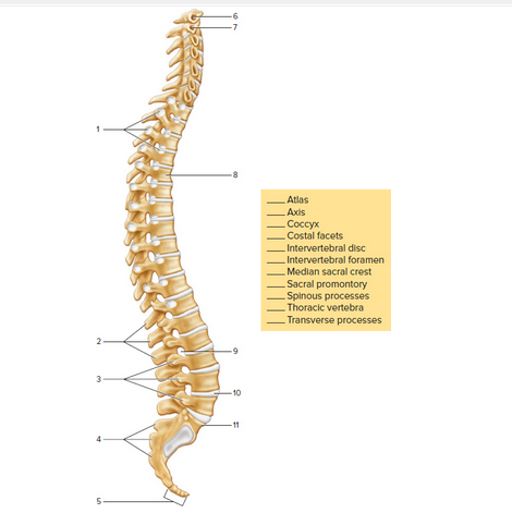
Expert Solution & Answer
Want to see the full answer?
Check out a sample textbook solution
Students have asked these similar questions
Figure 7b.7. The left orbit, anterior view. (a) Structures of the orbit. (b) Photograph of the orbit to be labeled.
5.
2. SUPIOOYBHA maYgin
* Z49omatlC
1acrimal BOne
OPTIC Canal
7. SUPEKIOK OPDilal riEsuet
पाम्बनाप .
6.
When the head is moved from side to side, the first vertebra pivots around the _______________________ of thesecond vertebra.
From The anatomical position, the ulna is more lateral than the radius.
Chapter 15 Solutions
Laboratory Manual For Human Anatomy & Physiology
Ch. 15 - The most superior bone of the vertebral column is...Ch. 15 - The vertebral column possesses four curvatures....Ch. 15 - Humans have ___________ pairs of true ribs. two...Ch. 15 - The _________ ribs do not have costal cartilage...Ch. 15 - Humans possess ____________ cervical vertebrae....Ch. 15 - The superior end of the sacrum articulates with...Ch. 15 - The anterior (sternal) end of a rib articulates...Ch. 15 - All cervical, thoracic, and lumbar vertebrae...Ch. 15 - A feature of the second cervical vertebra is the...Ch. 15 - Note the four curvatures of the vertebral column....
Ch. 15 - The vertebral column encloses and protects the...Ch. 15 - The vertebral column extends from the skull to the...Ch. 15 - The seventh cervical vertebra is called the...Ch. 15 - The _____________________ of the vertebrae support...Ch. 15 - The __________ separate adjacent vertebrae, and...Ch. 15 - The intervertebral foramina provide passageways...Ch. 15 - Transverse foramina of _____________ vertebrae...Ch. 15 - The first vertebra also is called the...Ch. 15 - When the head is moved from side to side, the...Ch. 15 - The __________ vertebrae have the largest and...Ch. 15 - The typical number ofvertebrae that fuse in the...Ch. 15 - FIGURE 15.8 Label the bones and features of a...Ch. 15 - FIGURE 15.9 Identity the bones and features...Ch. 15 - An abnormal lateral curvature of the spine is...Ch. 15 - The manubrium, body, and xiphoid process form a...Ch. 15 - The last two pairs of fibs that have no...Ch. 15 - There are _____________ pairs of true ribs.Ch. 15 - Costal are composed of __________ tissue.Ch. 15 - The manubriunarticulates with _________ on its...Ch. 15 - List three general functions ofthe thoracic cage.Ch. 15 - The sternal angle indicates the location of the...Ch. 15 - FIGURE 15.11 Label the bones and features of the...
Additional Science Textbook Solutions
Find more solutions based on key concepts
Define histology.
Fundamentals of Anatomy & Physiology (11th Edition)
More than one choice may apply. Using the terms listed below, fill in the blank with the proper term. anterior ...
Essentials of Human Anatomy & Physiology (11th Edition)
Describe the evolution of mammals, tracing their synapsid lineage from early amniote ancestors to true mammals....
Loose Leaf For Integrated Principles Of Zoology
Nursing Student with Neuropathic Pain
Tamara Costa broke her right tibia and has undergone two separate surger...
Human Anatomy & Physiology (11th Edition)
Describe the evolution of mammals, tracing their synapsid lineage from early amniote ancestors to true mammals....
LooseLeaf for Integrated Principles of Zoology
Knowledge Booster


Learn more about
Need a deep-dive on the concept behind this application? Look no further. Learn more about this topic, biology and related others by exploring similar questions and additional content below.Similar questions
- The image shows the last 2 thoracic vertebrae of DOG (T12,T13) in lateral view. Find and label the following: Arch Body Spinous process Caudal articular process Cranial articular process Mammillary process Accessory process Fovea of transverse process Cranial costal fovea Lamina Pediclearrow_forwardLabel the numbered parts of the vertebrae. (Dorsal view of the vertebrae)arrow_forward2 3 4 Figure 31.7th cervical vertebra, superior view. This bone shows the basic parts of a vertebra. 5 6 7 8arrow_forward
- Observe the bones in Figure (a) and (b). Identify the child’s hand and the adult’s hand. (a) _________________________________________ (b) ____________________________________________arrow_forwardLumbar Vertebra Observe a lumbar vertebra and using Figure 7B.17 as a reference, identify its structures. Then fill in the blanks below Figure 7B.17 to label the photographs. PASSPO ASAR 4 Intervertebral notch • on all vertebrae below the pedicle • nerves and blood vessels to and from spinal cord pass through these spaces Inferior articular facet • on all vertebrae • articulates with the superior articular facet of the vertebrae below it 3. 4. tioonanig 2. Transverse foramen seen only in cervical vertebrae O bluedoor, LLC protects vertebral arteries going to and veins coming from the brain 4. 5. Figure 7b.15. A typical cervical vertebra, superior view. Photograph of the cervical vertebra to be labeled. 1. 5. 3.arrow_forwardPlease label again the skull using the following terms: A colored dot at the end of a leader line indicates a bone. Leader line without a colored dot indicate bone markings. (Note: vomer, sphenoid bone and zygomatic bone will each be labeled twice) 1. alveolar processes 2. carotid canal 3. ethmoid bone (perpendicular plate) 4. external occipital protuberance 5. foramen lacerum 6. foramen magnum 7. foramen ovale 8. frontal bone 9. glabella 10. incisive fossa 11. inferior nasal concha 12. inferior orbital fissure 13. infraorbital foramen 14. jugular foramen 15. lacrimal bone 16. mandible 17. mandibular fossa 18. mandibular symphysis 19. mastoid process 20. maxilla 21. mental foramen 22. nasal bone 23. occipital bone 24. occipital condyle 25. palatine bone 26. palatine process of maxilla 27. parietal bone 28. sphenoid bone 29. styloid process 30. stylomastoid foramen 31. superior orbital fissure 32. supraorbital foramen 33. temporal bone 34. vomer bone 35. zygomatic bone 36. zygomatic…arrow_forward
- Using the image provided, identify the vertebra that the arrow is pointing to. O T11 OT12 O L1 O L2 OL3arrow_forwardUse the labels from figure 13.1 and color in the bones of the upper extremity in this anterior view. The inset is a posterior view of the elbow jointarrow_forwardSpecimen: Chicken Bones Lumbar and sacral vertebrae: There are several bones in synsacrum (made up of thoracic, lumbar, and sacral vertebrae). Describe the synsacrum in your specimen.arrow_forward
arrow_back_ios
SEE MORE QUESTIONS
arrow_forward_ios
Recommended textbooks for you

The Skeletal System; Author: Professor Dave Explains;https://www.youtube.com/watch?v=f-FF7Qigd3U;License: Standard YouTube License, CC-BY