Basic Clinical Laboratory Techniques 6E
6th Edition
ISBN: 9781133893943
Author: ESTRIDGE
Publisher: Cengage
expand_more
expand_more
format_list_bulleted
Question
thumb_up100%

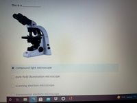
Transcribed Image Text:This is a
compound light microscope
O dark-field illumination microscope
O scanning electron microscope
transmission electron microscope
71°F Sunny
立
Expert Solution
arrow_forward
Step 1
The cell is very small in size. It is unable to see by naked eyes. So, scientists used microscopes to study them. The microscope is the instrument that helps to magnify small things. With the help of a microscope, we can study bacteria, viruses, cells, etc. Scientists have been able to answer many questions through microscopic observations of cells. We are able to see cells move and their tiny components work together. Without the microscope, we would not understand many of the inner workings of cells. The Cell Theory emerged from early work with microscopes.
Step by stepSolved in 2 steps

Knowledge Booster
Learn more about
Need a deep-dive on the concept behind this application? Look no further. Learn more about this topic, biology and related others by exploring similar questions and additional content below.Similar questions
- What is the highest magnification of the following microscopes?Bright fieldDark fieldPhase-contrastFluorescenceConfocalScanning EMTransmission EMScanned-Probearrow_forwardAll of the following are types of light microscopes except electron phase-contrast Obright-field O darkfieldarrow_forwardYou are in charge of buying new microscopes for your company, and you have been comparing different types of microscopes. One important property for microscopy is the resolution, which measures the ability to distinguish two small objects that are close together. Rank the following list of microscopes from lowest to highest resolution. Microscope List (5 items) (Drag and drop into the appropriate area) Transmission electron microscope (uses high energy electron beam) X-ray microscope (uses x-ray absorption to image sample, range 0.01-10 nm) Confocal fluorescence microscope (uses a blue laser as a light source, 488 nm) Level of Resolution 2 Lowestarrow_forward
- 2 bright-fleld scanning electron microscopy microscopy microscopy 3 interference 4 microscopy microscopyarrow_forwardWhich of the following is never useful for observing living cells? O phase-contrast microscope scanning electron microscope O darkfield microscope O brightficld microscope confocal microscopearrow_forwardIdentify and indicate the function of the parts of the dissecting microscope A B C D -E B Farrow_forward
- In order to visualize the fine structure of viruses and cytoskeletal filaments at 10-25 nanometers in diameter the type of microscopy that would be most effective is O standard light microscopy O phase-contrast light microscopy transmission electron microscopy O darkfield light microscopy O differential-interference microscopyarrow_forwardWhich microscope achieves the highest magnification and greatest resolution? phase-contrast microscope compound light microscope darkfield microscope electron microscope O fluorescence microscopearrow_forwardHelp witharrow_forward
- For each type of microscopy, know how each is used and the type of image produced. kEEP IT SHORT AND SWEET Light microscope – Transmission electron microscope - Scanning electron microscope –arrow_forwardAn object found with the 10x objective of the microscope may be viewed with the 40x objective without having to re-focus. This property of the microscope is called: O Confocal O Bifocal Binocular d Parfocalarrow_forwardWithin the microscope image below, identify the type of microscopy used by dragging the correct label to the target in the image. A Electron 10 μm Q microscope B Fluorescence microscopearrow_forward
arrow_back_ios
SEE MORE QUESTIONS
arrow_forward_ios
Recommended textbooks for you
