Human Anatomy & Physiology (11th Edition)
11th Edition
ISBN: 9780134580999
Author: Elaine N. Marieb, Katja N. Hoehn
Publisher: PEARSON
expand_more
expand_more
format_list_bulleted
Question
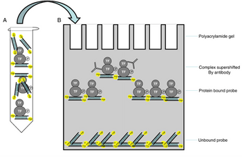
The schematic on the right is for which molecular biology method?
What information does this method reveal?

Transcribed Image Text:**Transcription and Explanation for an Educational Website**
**Title: Understanding Gel Electrophoresis in Protein-DNA Interactions**
**Diagram Explanation:**
The image illustrates the process of gel electrophoresis used to study protein-DNA interactions, particularly focusing on the binding of transcription factors (TF) to DNA probes. The diagram is divided into two main parts:
**A: Mixture Preparation**
- This section shows a test tube containing a mixture of DNA probes and transcription factors (TF).
- The DNA probes are represented as elongated rectangles, some of which have transcription factors bound to them.
- The free probes are depicted as having no transcription factor attached.
**B: Gel Electrophoresis Process**
- The polyacrylamide gel is used to separate different complexes based on size and charge.
- The gel contains several wells (topmost) where the prepared samples are loaded.
- As the samples migrate through the gel:
1. **Complex Supershifted By Antibody:**
- These are shown as the slowest migrating bands in the gel.
- They consist of DNA probes bound to transcription factors, which are further bound by specific antibodies, increasing the molecular weight and causing a supershift.
2. **Protein Bound Probe:**
- This section shows DNA probes bound to transcription factors.
- These complexes migrate more slowly than unbound probes but faster than complex supershifted by antibody.
3. **Unbound Probe:**
- Represented at the bottom, these probes migrate the fastest through the gel.
- They indicate the portion of DNA probes that are not bound by any transcription factor.
**Conclusion:**
This diagram provides a visual representation of how gel electrophoresis can be used to analyze the binding interactions between proteins (such as transcription factors) and DNA. By comparing the migration patterns of different complexes, researchers can infer the presence and strength of protein-DNA interactions.
Expert Solution
This question has been solved!
Explore an expertly crafted, step-by-step solution for a thorough understanding of key concepts.
This is a popular solution
Trending nowThis is a popular solution!
Step by stepSolved in 3 steps

Knowledge Booster
Learn more about
Need a deep-dive on the concept behind this application? Look no further. Learn more about this topic, biology and related others by exploring similar questions and additional content below.Similar questions
- Gel Electrophoresis is used in many different forms to learn about DNA, RNA or proteins. Research one laboratory method or technique that uses DNA electrophoresis in order to learn more or make determinations about DNA, RNA or Proteins. Name of technique or method Brief description of what the electrophoresis results are used for in the method. Include a link to your resources Include an image of the results or technique you describearrow_forwardGenetic information from the Human Genome Project may be used to develop screening procedures which could be used by insurance companies or employers. Discuss the possible implications of the use of genetic information for this type of screeningarrow_forwardWhich lab technique would you use for each of the following: a.Break Open Cell Membrane b. Remove Cell Debris c.Amplify Region of Interest d. Analyze DNA for SNP Variationarrow_forward
- What are the reasons why there needs to be more than 10 cycles in the PCR process? Name 2 reasons.arrow_forwardWhat is the role of streptomycin in CRISPR experiment? What biochemical changes (DNA, protein) occurred in those cells in which CRISPR worked?arrow_forwardYour colleague wants to align the complete sequence of the collagen gene from humans to the homologous complete gene in rats. She can't decide if she should use the alignment program Needle or Water, and turns to you for help. Does it make a difference which program she uses, what advice would you give her?arrow_forward
- After running a qPCR experiment, we will have graphs showing the amount of fluorescence detected by the digital camera compared to the number of PCR cycles run. Suppose you see the following graph output by the qPCR machine: Relative Fluoresence 3.0 2.5 2.0 1.5 1.0 0.5 0.0 0 10 20 30 40 50 Cycles Which curve (blue, red, or green) represents a sample with the smallest amount of mRNA present? Why? Be sure to discuss Ct values in your answer.arrow_forwardSelect all that would be true if I had a nonsense mutation in an exon of a gene: The nonsense mutant allele would be the same size as wildtype by PCR-electrophoresis The nonsense mutant protein would be the same size by Western as the wildtype protein The nonsense mutant allele would be a different size compared to wildtype by PCR- electrophoresis The nonsense mutant protein would be a different size by Western compared to the wildtype proteinarrow_forwardWhat is the program Finch TV used for?arrow_forward
- Which of the following technologies would be useful to assess natural patterns of mRNA abundance in the brain of a reptile species that has never been the focus of genetic research before? Group of answer choices DNA microarray RNA-sequencing with Illumina DNA-sequencing with Illumina RNA microarrayarrow_forwardWhat is the Lineweaver-Burk plot and what does it tell us?arrow_forwardWhat are some of the technical challenges of cloning a mammoth? Check all that are true Ancient mammoth DNA has degraded so it is hard to know the complete genome sequence Mammoth gestation time is likely too short to allowing cloning A living surrogate mother would have to be used, which could pose problems since the closest relative to mammoths is endangered elephants A living oocyte would have to be obtained from an extant species, which could pose problems since the closest relative to mammoths is endangered elephants Mammoths lived so long ago that they used a different genetic code than modern animals There are no enzymes in existence that could ligate together mammoth DNA sequence with elephant sequence Mammoths probably had egg shells, which would be hard to penetrate with a needlearrow_forward
arrow_back_ios
SEE MORE QUESTIONS
arrow_forward_ios
Recommended textbooks for you
 Human Anatomy & Physiology (11th Edition)BiologyISBN:9780134580999Author:Elaine N. Marieb, Katja N. HoehnPublisher:PEARSON
 Biology 2eBiologyISBN:9781947172517Author:Matthew Douglas, Jung Choi, Mary Ann ClarkPublisher:OpenStax
 Anatomy & PhysiologyBiologyISBN:9781259398629Author:McKinley, Michael P., O'loughlin, Valerie Dean, Bidle, Theresa StouterPublisher:Mcgraw Hill Education,
 Molecular Biology of the Cell (Sixth Edition)BiologyISBN:9780815344322Author:Bruce Alberts, Alexander D. Johnson, Julian Lewis, David Morgan, Martin Raff, Keith Roberts, Peter WalterPublisher:W. W. Norton & Company
 Laboratory Manual For Human Anatomy & PhysiologyBiologyISBN:9781260159363Author:Martin, Terry R., Prentice-craver, CynthiaPublisher:McGraw-Hill Publishing Co.
 Inquiry Into Life (16th Edition)BiologyISBN:9781260231700Author:Sylvia S. Mader, Michael WindelspechtPublisher:McGraw Hill Education

Human Anatomy & Physiology (11th Edition)
Biology
ISBN:9780134580999
Author:Elaine N. Marieb, Katja N. Hoehn
Publisher:PEARSON

Biology 2e
Biology
ISBN:9781947172517
Author:Matthew Douglas, Jung Choi, Mary Ann Clark
Publisher:OpenStax

Anatomy & Physiology
Biology
ISBN:9781259398629
Author:McKinley, Michael P., O'loughlin, Valerie Dean, Bidle, Theresa Stouter
Publisher:Mcgraw Hill Education,

Molecular Biology of the Cell (Sixth Edition)
Biology
ISBN:9780815344322
Author:Bruce Alberts, Alexander D. Johnson, Julian Lewis, David Morgan, Martin Raff, Keith Roberts, Peter Walter
Publisher:W. W. Norton & Company

Laboratory Manual For Human Anatomy & Physiology
Biology
ISBN:9781260159363
Author:Martin, Terry R., Prentice-craver, Cynthia
Publisher:McGraw-Hill Publishing Co.

Inquiry Into Life (16th Edition)
Biology
ISBN:9781260231700
Author:Sylvia S. Mader, Michael Windelspecht
Publisher:McGraw Hill Education