Human Anatomy & Physiology (11th Edition)
11th Edition
ISBN: 9780134580999
Author: Elaine N. Marieb, Katja N. Hoehn
Publisher: PEARSON
expand_more
expand_more
format_list_bulleted
Question
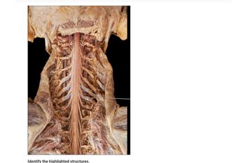
Ventral root of ganglia?

Transcribed Image Text:**Image Description and Analysis**
This image shows a detailed view of the central nervous system, specifically highlighting the spinal cord and associated structures within the thoracic region of a human body dissection.
**Key Features:**
1. **Spinal Cord:**
- Located centrally, the spinal cord extends vertically through the vertebral column.
- It is protected by vertebrae and is part of the central nervous system.
2. **Nerve Roots:**
- Emanating laterally from the spinal cord, the nerve roots appear in pairs.
- These are responsible for transmitting signals between the spinal cord and the rest of the body.
3. **Vertebrae:**
- The bony structures surrounding the spinal cord, providing protection and support.
- Vertebrae are identifiable around the spinal cord.
4. **Intercostal Nerves:**
- Running in between the ribs, these are visible diverging from the spinal nerves.
5. **Highlighted Structures:**
- The image includes blue lines indicating specific highlighted structures, which likely reference targeted nerves or vessels for educational focus.
This image is often used in medical and anatomy education to illustrate the relationship between the spinal cord, nerves, and surrounding structures. Understanding this layout is crucial for comprehending the complex communication network within the human body. Students could be asked to identify the highlighted structures to test their knowledge of spinal anatomy.
**Instruction:**
At the bottom of the image, viewers are prompted with the text:
- "Identify the highlighted structures."
This suggests an interactive element for educational purposes, encouraging users to engage with the material by identifying key anatomical components shown in the image.
Expert Solution
This question has been solved!
Explore an expertly crafted, step-by-step solution for a thorough understanding of key concepts.
This is a popular solution
Trending nowThis is a popular solution!
Step by stepSolved in 3 steps

Follow-up Questions
Read through expert solutions to related follow-up questions below.
Follow-up Question
"Spinal Nerve-Rootlets" sound acceptable, this is being received during lab practical online work.
Solution
by Bartleby Expert
Follow-up Questions
Read through expert solutions to related follow-up questions below.
Follow-up Question
"Spinal Nerve-Rootlets" sound acceptable, this is being received during lab practical online work.
Solution
by Bartleby Expert
Knowledge Booster
Similar questions
- Possible Explanation to Neuroscience Question: Given that both B and C appear to be in the gray matter, and the axons of descending tracts like the vestibulospinal tract would be found in the white matter, the correct location would likely be one of the areas in the white matter. From the diagram, areas A, D, and E are left. Typically, descending motor tracts like the vestibulospinal tract run in the anterolateral part of the white matter. Given this, area D would be a likely location for the axons of the vestibulospinal tract. So, the answer would be: Darrow_forwardQuestion: Label the white matter, gray matter, and a neuron cell body.arrow_forward
Recommended textbooks for you
 Human Anatomy & Physiology (11th Edition)Anatomy and PhysiologyISBN:9780134580999Author:Elaine N. Marieb, Katja N. HoehnPublisher:PEARSON
 Anatomy & PhysiologyAnatomy and PhysiologyISBN:9781259398629Author:McKinley, Michael P., O'loughlin, Valerie Dean, Bidle, Theresa StouterPublisher:Mcgraw Hill Education,
 Human AnatomyAnatomy and PhysiologyISBN:9780135168059Author:Marieb, Elaine Nicpon, Brady, Patricia, Mallatt, JonPublisher:Pearson Education, Inc.,
 Anatomy & Physiology: An Integrative ApproachAnatomy and PhysiologyISBN:9780078024283Author:Michael McKinley Dr., Valerie O'Loughlin, Theresa BidlePublisher:McGraw-Hill Education
 Human Anatomy & Physiology (Marieb, Human Anatomy...Anatomy and PhysiologyISBN:9780321927040Author:Elaine N. Marieb, Katja HoehnPublisher:PEARSON

Human Anatomy & Physiology (11th Edition)
Anatomy and Physiology
ISBN:9780134580999
Author:Elaine N. Marieb, Katja N. Hoehn
Publisher:PEARSON

Anatomy & Physiology
Anatomy and Physiology
ISBN:9781259398629
Author:McKinley, Michael P., O'loughlin, Valerie Dean, Bidle, Theresa Stouter
Publisher:Mcgraw Hill Education,

Human Anatomy
Anatomy and Physiology
ISBN:9780135168059
Author:Marieb, Elaine Nicpon, Brady, Patricia, Mallatt, Jon
Publisher:Pearson Education, Inc.,

Anatomy & Physiology: An Integrative Approach
Anatomy and Physiology
ISBN:9780078024283
Author:Michael McKinley Dr., Valerie O'Loughlin, Theresa Bidle
Publisher:McGraw-Hill Education

Human Anatomy & Physiology (Marieb, Human Anatomy...
Anatomy and Physiology
ISBN:9780321927040
Author:Elaine N. Marieb, Katja Hoehn
Publisher:PEARSON