Human Anatomy & Physiology (11th Edition)
11th Edition
ISBN: 9780134580999
Author: Elaine N. Marieb, Katja N. Hoehn
Publisher: PEARSON
expand_more
expand_more
format_list_bulleted
Concept explainers
Question
thumb_up100%
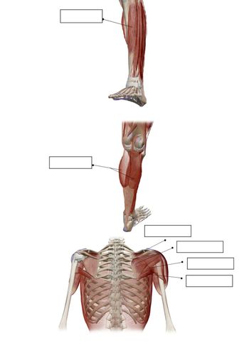
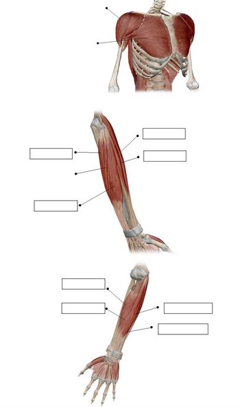
Fill in the blanks in the image given:

Transcribed Image Text:The image contains diagrams focusing on different muscle groups within the human body, specifically in the leg and shoulder areas. Each section of the image has labels indicating specific muscles.
### Leg Muscles:
1. **Quadriceps (front of thigh)**: This group of muscles is responsible for extending the knee and is located in the front part of the thigh.
2. **Gastrocnemius (calf muscle)**: Located at the back of the lower leg, this muscle is involved in plantar flexing the foot at the ankle joint and flexing the leg at the knee joint.
### Shoulder Muscles:
1. **Deltoid**: A rounded, triangular muscle responsible for lifting the arm and giving the shoulder its range of motion.
2. **Trapezius**: Extending over the back of the neck and shoulders, this muscle facilitates neck and shoulder movement.
3. **Latissimus Dorsi**: A large, flat muscle on the back that helps with arm movement and rotation, often contributing to shoulder and back strength.
These anatomical illustrations highlight the muscle positioning and potential actions they facilitate within the body, providing a clear visual understanding for educational purposes.

Transcribed Image Text:The image consists of anatomical diagrams highlighting various muscles of the upper body and arm. The diagrams are labeled with placeholders for identifying specific muscles. Here's a detailed breakdown:
1. **Shoulder Muscles**:
- **Upper Diagram**: Displays the front view of the chest and shoulder. Muscles like the pectoralis major and deltoid are shown. Blank labels are present for identifying each muscle group.
2. **Upper Arm Muscles**:
- **Middle Diagram**: Focuses on the upper arm, displaying both anterior (front) muscles such as the biceps brachii, and possibly posterior muscles like the triceps brachii. It includes several blank labels indicating different muscle regions.
3. **Forearm Muscles**:
- **Lower Diagram**: Illustrates the muscles of the forearm and hand. Visible muscles might include the flexor and extensor groups, with more blank labels pointing out various muscle areas.
This detailed visual aid serves as an educational tool for identifying major muscle groups involved in upper body function.
Expert Solution
This question has been solved!
Explore an expertly crafted, step-by-step solution for a thorough understanding of key concepts.
Step by stepSolved in 3 steps with 2 images

Knowledge Booster
Learn more about
Need a deep-dive on the concept behind this application? Look no further. Learn more about this topic, biology and related others by exploring similar questions and additional content below.Similar questions
arrow_back_ios
arrow_forward_ios
Recommended textbooks for you
 Human Anatomy & Physiology (11th Edition)BiologyISBN:9780134580999Author:Elaine N. Marieb, Katja N. HoehnPublisher:PEARSON
 Biology 2eBiologyISBN:9781947172517Author:Matthew Douglas, Jung Choi, Mary Ann ClarkPublisher:OpenStax
 Anatomy & PhysiologyBiologyISBN:9781259398629Author:McKinley, Michael P., O'loughlin, Valerie Dean, Bidle, Theresa StouterPublisher:Mcgraw Hill Education,
 Molecular Biology of the Cell (Sixth Edition)BiologyISBN:9780815344322Author:Bruce Alberts, Alexander D. Johnson, Julian Lewis, David Morgan, Martin Raff, Keith Roberts, Peter WalterPublisher:W. W. Norton & Company
 Laboratory Manual For Human Anatomy & PhysiologyBiologyISBN:9781260159363Author:Martin, Terry R., Prentice-craver, CynthiaPublisher:McGraw-Hill Publishing Co.
 Inquiry Into Life (16th Edition)BiologyISBN:9781260231700Author:Sylvia S. Mader, Michael WindelspechtPublisher:McGraw Hill Education

Human Anatomy & Physiology (11th Edition)
Biology
ISBN:9780134580999
Author:Elaine N. Marieb, Katja N. Hoehn
Publisher:PEARSON

Biology 2e
Biology
ISBN:9781947172517
Author:Matthew Douglas, Jung Choi, Mary Ann Clark
Publisher:OpenStax

Anatomy & Physiology
Biology
ISBN:9781259398629
Author:McKinley, Michael P., O'loughlin, Valerie Dean, Bidle, Theresa Stouter
Publisher:Mcgraw Hill Education,

Molecular Biology of the Cell (Sixth Edition)
Biology
ISBN:9780815344322
Author:Bruce Alberts, Alexander D. Johnson, Julian Lewis, David Morgan, Martin Raff, Keith Roberts, Peter Walter
Publisher:W. W. Norton & Company

Laboratory Manual For Human Anatomy & Physiology
Biology
ISBN:9781260159363
Author:Martin, Terry R., Prentice-craver, Cynthia
Publisher:McGraw-Hill Publishing Co.

Inquiry Into Life (16th Edition)
Biology
ISBN:9781260231700
Author:Sylvia S. Mader, Michael Windelspecht
Publisher:McGraw Hill Education