Human Anatomy & Physiology (11th Edition)
11th Edition
ISBN: 9780134580999
Author: Elaine N. Marieb, Katja N. Hoehn
Publisher: PEARSON
expand_more
expand_more
format_list_bulleted
Concept explainers
Question
give answer asap please.

Transcribed Image Text:**Ultrasonography and Genetic Analysis in Prenatal Diagnosis**
*Fetal Abnormalities Detected in Early Pregnancy*
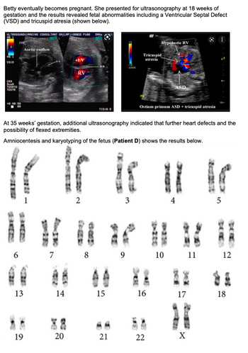
Betty, after becoming pregnant, underwent an ultrasonography at 18 weeks of gestation. The ultrasound results revealed significant fetal abnormalities, specifically a Ventricular Septal Defect (VSD) and tricuspid atresia.
**Ultrasound Analysis:**
- The left image displays two main structures: the left ventricle (LV) and the right ventricle (RV). There is an indication of a defect in the ventricular septum (VSD), which separates the left and right ventricles.
- The right image highlights additional abnormalities such as a hypoplastic right ventricle (RV) and tricuspid atresia. There is an indication of an atrial septal defect (ASD), specifically the ostium primum type, associated with tricuspid atresia.
*Further Findings Later in Pregnancy*
At 35 weeks of gestation, a subsequent ultrasound suggested additional heart defects and the possibility of flexed extremities.
*Genetic Analysis Through Amniocentesis*
Amniocentesis and karyotyping were performed on the fetus (referred to as Patient D), which produced the following chromosomal results:
**Karyotype Description:**
- The image depicts the chromosomes of Patient D, arranged in pairs from 1 to 22, along with the sex chromosomes, X.
- The karyotype appears to consist of normal pairs, indicating no apparent chromosomal abnormalities.
This case highlights the importance of comprehensive prenatal testing, combining ultrasonography with genetic analysis for early detection and management of fetal conditions.

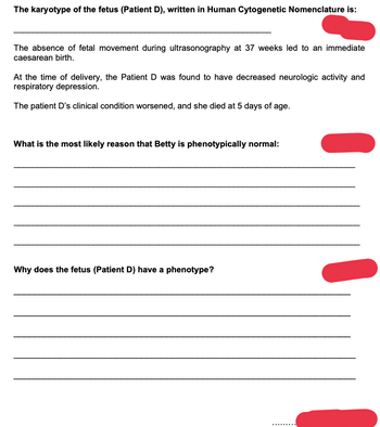
Transcribed Image Text:**The karyotype of the fetus (Patient D), written in Human Cytogenetic Nomenclature is:**
_____________________________________________________________
The absence of fetal movement during ultrasonography at 37 weeks led to an immediate caesarean birth.
At the time of delivery, the Patient D was found to have decreased neurologic activity and respiratory depression.
The patient D's clinical condition worsened, and she died at 5 days of age.
**What is the most likely reason that Betty is phenotypically normal:**
_____________________________________________________________
_____________________________________________________________
_____________________________________________________________
_____________________________________________________________
_____________________________________________________________
**Why does the fetus (Patient D) have a phenotype?**
_____________________________________________________________
_____________________________________________________________
_____________________________________________________________
_____________________________________________________________
_____________________________________________________________
Expert Solution
This question has been solved!
Explore an expertly crafted, step-by-step solution for a thorough understanding of key concepts.
Step by stepSolved in 2 steps

Knowledge Booster
Learn more about
Need a deep-dive on the concept behind this application? Look no further. Learn more about this topic, biology and related others by exploring similar questions and additional content below.Similar questions
- Part A Thyroxine: (Select all correct answers) is produced inside the follicular cells in response to TSH is produced within the colloid regardless of the presence of TSH O is the combination of DIT + DIT contains 3 atoms of iodine contains 4 atoms of iodine Submit Request Answer rovide Feedbackarrow_forwardAnswer the question please.arrow_forwardLabel the name for each numberarrow_forward
- Write a summary on Hormone? Or write a short note on hormone? Please write at your own words. Answer should be specific (5-6)lines.arrow_forwardI need help pleasearrow_forwardCan someone help me answer questions #5-8, please? Thank you!! 5. A) Define goiter. B) Explain, in terms of the thyroid gland, the reason why a goiter could be a sign for hyperthyroidism and why a goiter could be a sign for hypothyroidism. 6. A) Name two problems associated with hyperparathyroidism. B) Explain, in cellular terms, why each of those problems exist. C) What is a problem associated with hypoparathyroidism? D) Explain, in cellular terms, why that problem (stated in C)) exists. 7. A) Differentiate between Type I and Type II diabetes mellitus. B) Describe two major effects of diabetes on the cardiovascular system. C) Differentiate between diabetic versus insulin shock and describe how each would be treated. D) Consider the effects of insulin on blood levels of glucose and amino acids. Now explain why you should eat carbohydrates when eating a beefsteak (besides being a 'balanced diet'). 8. A) Define Addison's Disease. B) Describe a mineralocorticoid-based problem and a…arrow_forward
- Part I – SymptomsCallie was 26 years old when she opened a bakery called “Callie’s Cupcakes” in downtown San Francisco with herf ancé, Jeremy. Despite the competitive market, her business was booming; everyone loved the clever recipes and thetrendy atmosphere. Between running their fast-growing business and planning for their wedding, Callie hadn’t beenable to keep to her usual eight hours of sleep a night. Although she had always lived a very healthy lifestyle, exercisingdaily and eating healthy, she just hadn’t been feeling herself lately. She was tired all the time, had dif culty breathing,felt stressed, coughed up sputum, consistently ran a low-grade fever, and had lost weight as her appetite decreased.None of these symptoms alone had been particularly alarming so she had put of seeing her physician for a few weeks.Questions1. What are Callie’s symptoms? List all that were mentioned.2. Based on the symptoms presented, what are three possible respiratory infectious diseases Callie…arrow_forwardplease show on steparrow_forwarddiscuss two disease processes of the endocrine or nervous system that are most interesting to you. Why are you interested in these disease processes? Maximum of 300 words thanksarrow_forward
arrow_back_ios
SEE MORE QUESTIONS
arrow_forward_ios
Recommended textbooks for you
 Human Anatomy & Physiology (11th Edition)BiologyISBN:9780134580999Author:Elaine N. Marieb, Katja N. HoehnPublisher:PEARSON
 Biology 2eBiologyISBN:9781947172517Author:Matthew Douglas, Jung Choi, Mary Ann ClarkPublisher:OpenStax
 Anatomy & PhysiologyBiologyISBN:9781259398629Author:McKinley, Michael P., O'loughlin, Valerie Dean, Bidle, Theresa StouterPublisher:Mcgraw Hill Education,
 Molecular Biology of the Cell (Sixth Edition)BiologyISBN:9780815344322Author:Bruce Alberts, Alexander D. Johnson, Julian Lewis, David Morgan, Martin Raff, Keith Roberts, Peter WalterPublisher:W. W. Norton & Company
 Laboratory Manual For Human Anatomy & PhysiologyBiologyISBN:9781260159363Author:Martin, Terry R., Prentice-craver, CynthiaPublisher:McGraw-Hill Publishing Co.
 Inquiry Into Life (16th Edition)BiologyISBN:9781260231700Author:Sylvia S. Mader, Michael WindelspechtPublisher:McGraw Hill Education

Human Anatomy & Physiology (11th Edition)
Biology
ISBN:9780134580999
Author:Elaine N. Marieb, Katja N. Hoehn
Publisher:PEARSON

Biology 2e
Biology
ISBN:9781947172517
Author:Matthew Douglas, Jung Choi, Mary Ann Clark
Publisher:OpenStax

Anatomy & Physiology
Biology
ISBN:9781259398629
Author:McKinley, Michael P., O'loughlin, Valerie Dean, Bidle, Theresa Stouter
Publisher:Mcgraw Hill Education,

Molecular Biology of the Cell (Sixth Edition)
Biology
ISBN:9780815344322
Author:Bruce Alberts, Alexander D. Johnson, Julian Lewis, David Morgan, Martin Raff, Keith Roberts, Peter Walter
Publisher:W. W. Norton & Company

Laboratory Manual For Human Anatomy & Physiology
Biology
ISBN:9781260159363
Author:Martin, Terry R., Prentice-craver, Cynthia
Publisher:McGraw-Hill Publishing Co.

Inquiry Into Life (16th Edition)
Biology
ISBN:9781260231700
Author:Sylvia S. Mader, Michael Windelspecht
Publisher:McGraw Hill Education