Human Anatomy & Physiology (11th Edition)
11th Edition
ISBN: 9780134580999
Author: Elaine N. Marieb, Katja N. Hoehn
Publisher: PEARSON
expand_more
expand_more
format_list_bulleted
Concept explainers
Question
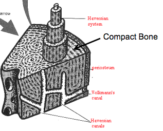
Attached is a compact bone of a femur.
What would be the simplest form of the elastic compliance matrix for the compact bone in the femur.
What aspects of the bone structure do you base your answer on?

Transcribed Image Text:arrow
Haversian
system
Compact Bone
periosteum
Vollmann's
canal
Haversian
canals
Expert Solution
This question has been solved!
Explore an expertly crafted, step-by-step solution for a thorough understanding of key concepts.
This is a popular solution
Trending nowThis is a popular solution!
Step by stepSolved in 2 steps

Knowledge Booster
Learn more about
Need a deep-dive on the concept behind this application? Look no further. Learn more about this topic, biology and related others by exploring similar questions and additional content below.Similar questions
- Label the following parts of the compact bone classroom models. Be sure to identify the parts on the in-class models too. Lacunae, Circumferential lamellae, Canaliculi, Perforating canal, Osteon, Central canal (x2), Periosteum, Sharpey's (Perforating) fibers, Osteocytes, Concentric lamellae, and Interstitial lamellae (cavities which nouse osteocytes) 43 Osteon Model Draw the provided tissue image (or microscope slide) of compact osseous tissue and label the following parts: Central canal, osteocytes, lacunae, canaliculi, concentric lamellae, and interstitial lamellae.arrow_forwardBased on the information and considering significant figures, what is the length of the femur bone? 6 7 8 cm 8.3 cm O 8.3333 cm O 8 cm 8.33 cmarrow_forwardWe are focusing mainly on synovial joints, because this is the main type of joint that allows you to move your body. Using the diagram below, match the synovial joint structure with its description: -Periosteum E F A G D H- F V [ Choose ] A Friction-reducing hyaline cartilage that covers bone surfaces B Cavity filled with lubricating, nourishing, and shock-absorbing fluid Bands of dense regular connective tissue that connect muscle to bone and help stabilize joints Fluid-filled pocket that reduces friction between joint structures Bands of dense regular connective tissue that connect bones Cushions of fibrous cartilage that help guide joint movement E Protective outer wrapping made of dense irregular connective tissue One of the four body membranes; produces synovial fluid F G [ Choose ] [ Choose ]arrow_forward
- While playing on her set, 10 year old sally falls and breaks her right leg. At the emergency room, the doctors tells her parents that the proximal end of the tibia where the epiphysis meets the diaphysis is fractured. The fracture is properly set and eventually heals. During a routine physical when she is 18, sally learns that her right leg is 2 cm shorter that her left, probably because of her accident. What might account for this difference?arrow_forwardDual Energy Xray Absorptiometry (DEXA) is used to determine bone mineral density and the risk of osteoporosis. Which of the follow statements regarding DEXA is true? a T-score of -4.0 indicates healthy bone mineral density with no risk of fracture during a fall Osteoporotic bone develops in both trabecular and cortical bone The very high levels of radiation used in a DEXA scan make this is a dangerous procedure One of the most common sites to perform a DEXA scan is in the hip area Two of the above statements are truearrow_forwarda Parallel lamellae, endosteum, lines of stress. 8. Describe the roles played by OSTEOBLASTS, OSTEOCYTES and OSTEOCLASTS. 9. Name the major inorganic and organic components of bone matrix. Also, identify what would happen to bones if the organic component is removed. If the horganic component is removed? 10. Describe and contrast INTRAMEMBRANOUS and ENDOCHONDRAL OSSIFICATION.arrow_forward
- For this installment of your discussions please give us two main differences or elaborate on any you can think of between the two divisions of the skeletal system; from the most apparent to how these bones develop. Make your comments brief and succinct. If it seems as though the contributions are sounding similar or the same include a trivia like or little known fact about any one bone in the skeletal system. I will give you an example: The axial contains all bones within the medial aspect of the body. The appendicular incorporates the bones of the pectoral, pelvic girdles and extremities. The capitate is the largest bone of the carpal bones.arrow_forwardPlease answer the following question:arrow_forwardWhich of the following is/are true of bone? 1. chondrocytes present 2. low ground substance 3. high collagen fiber content 4. lacunae present 5. high reticular fiber content O 1, 2, 3, 4, 5 O 2, 3, 4, 5 O 2, 3, 4 O 1, 2, 3, 4 O 2,5arrow_forward
- In addition to the tuberosity, what other feature of the tibia can you use to help determine if it is from the left side or the right side of the body? What kind of joint (structurally and functionally) is the ankle joint? What is the common name for the calcaneus on the human body? How many phalanges are found in an entire human body? What bone does the acromion articulate with? How would you describe a notch? What bone articulates with the glenoid cavity of the scapula? What are the names acromion and sternal ends telling you? Is the glenoid a shallow or deep socket? How will this affect the stability and mobility of the joint? Which group of four muscles inserts on the greater and lesser tubercles?arrow_forwardThe Musculoskeletal System Name of Condition Which part of the system is affected? In what way? What causes this condition? What are the objective/subjective signs & symptoms? What is the usual treatment for this condition? What diagnostic tools would be used? Results? Fractures Osteoporosis Osteoarthritis Osteomyelitis Goutarrow_forwardThe hand and foot are structurally similar in many ways; they also show clear differences in structure related to their different functions. Describe the structural features of the foot that are clearly related to its weight-bearing and locomotory functions.arrow_forward
arrow_back_ios
SEE MORE QUESTIONS
arrow_forward_ios
Recommended textbooks for you
 Human Anatomy & Physiology (11th Edition)BiologyISBN:9780134580999Author:Elaine N. Marieb, Katja N. HoehnPublisher:PEARSON
 Biology 2eBiologyISBN:9781947172517Author:Matthew Douglas, Jung Choi, Mary Ann ClarkPublisher:OpenStax
 Anatomy & PhysiologyBiologyISBN:9781259398629Author:McKinley, Michael P., O'loughlin, Valerie Dean, Bidle, Theresa StouterPublisher:Mcgraw Hill Education,
 Molecular Biology of the Cell (Sixth Edition)BiologyISBN:9780815344322Author:Bruce Alberts, Alexander D. Johnson, Julian Lewis, David Morgan, Martin Raff, Keith Roberts, Peter WalterPublisher:W. W. Norton & Company
 Laboratory Manual For Human Anatomy & PhysiologyBiologyISBN:9781260159363Author:Martin, Terry R., Prentice-craver, CynthiaPublisher:McGraw-Hill Publishing Co.
 Inquiry Into Life (16th Edition)BiologyISBN:9781260231700Author:Sylvia S. Mader, Michael WindelspechtPublisher:McGraw Hill Education

Human Anatomy & Physiology (11th Edition)
Biology
ISBN:9780134580999
Author:Elaine N. Marieb, Katja N. Hoehn
Publisher:PEARSON

Biology 2e
Biology
ISBN:9781947172517
Author:Matthew Douglas, Jung Choi, Mary Ann Clark
Publisher:OpenStax

Anatomy & Physiology
Biology
ISBN:9781259398629
Author:McKinley, Michael P., O'loughlin, Valerie Dean, Bidle, Theresa Stouter
Publisher:Mcgraw Hill Education,

Molecular Biology of the Cell (Sixth Edition)
Biology
ISBN:9780815344322
Author:Bruce Alberts, Alexander D. Johnson, Julian Lewis, David Morgan, Martin Raff, Keith Roberts, Peter Walter
Publisher:W. W. Norton & Company

Laboratory Manual For Human Anatomy & Physiology
Biology
ISBN:9781260159363
Author:Martin, Terry R., Prentice-craver, Cynthia
Publisher:McGraw-Hill Publishing Co.

Inquiry Into Life (16th Edition)
Biology
ISBN:9781260231700
Author:Sylvia S. Mader, Michael Windelspecht
Publisher:McGraw Hill Education