Human Anatomy & Physiology (11th Edition)
11th Edition
ISBN: 9780134580999
Author: Elaine N. Marieb, Katja N. Hoehn
Publisher: PEARSON
expand_more
expand_more
format_list_bulleted
Concept explainers
Question

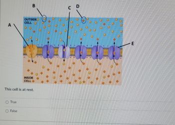
Transcribed Image Text:The image depicts a cross-section of a cell membrane illustrating the transport of ions across the membrane.
### Diagram Explanation:
- **Area Labelled A:** Indicates a channel in the membrane that facilitates active transport.
- **Area Labelled B:** Represents ions outside of the cell, specifically sodium ions (Na⁺), depicted as small yellow circles.
- **Area Labelled C:** Demonstrates ion channels embedded in the membrane, allowing for passive transport of ions.
- **Area Labelled D:** Shows potassium ions (K⁺) outside the cell, depicted as larger orange circles.
- **Area Labelled E:** Indicates the transport directions of ions through the channels.
**Membrane Structure:**
- **Outside Cell:** The region above the membrane, highlighted in blue, represents the extracellular space.
- **Inside Cell:** The region below the membrane, highlighted in beige, represents the intracellular space.
- **Ion Channels:** Shown as purple structures spanning the membrane, facilitating ion movement.
### True/False Question:
This cell is at rest.
- ○ True
- ○ False
Note that the cellular activity related to ion movement implies a dynamic equilibrium state rather than a complete rest state.
![**Action Potential Phases Matching Exercise**
In this exercise, you will match the different phases of an action potential depicted in the plot with their correct terms or descriptions. The graph illustrates the changes in membrane potential over time, marked from A to E, each representing a distinct phase in the action potential.
**Graph Description:**
- **Y-Axis (Membrane Potential):** Ranges from -70 mV to +50 mV.
- **X-Axis (Time):** Ranges from 0 to 4 units.
**Phases:**
- **A:** This phase begins at the resting membrane potential (-70 mV) and shows a slight initial increase.
- **B:** A rapid rise in membrane potential, indicating depolarization, reaching a peak of about +50 mV.
- **C:** A sharp decrease from the peak, representing repolarization back toward negative values.
- **D:** A slight overshoot beyond the resting potential, indicating hyperpolarization.
- **E:** Horizontal line indicating the return to the resting membrane potential.
**Matching Terms/Descriptions:**
- **A [] Choose**
- **B [] Choose**
- **C [] Choose**
- **D [] Choose**
- **E [] Choose**
Select the most appropriate term or description for each labeled phase in the provided dropdowns.](https://content.bartleby.com/qna-images/question/450044ef-db4b-4b73-a24e-98f3627ca204/9e7275a4-1130-48dd-b668-d66df3ef8ba5/g9li8_thumbnail.jpeg)
Transcribed Image Text:**Action Potential Phases Matching Exercise**
In this exercise, you will match the different phases of an action potential depicted in the plot with their correct terms or descriptions. The graph illustrates the changes in membrane potential over time, marked from A to E, each representing a distinct phase in the action potential.
**Graph Description:**
- **Y-Axis (Membrane Potential):** Ranges from -70 mV to +50 mV.
- **X-Axis (Time):** Ranges from 0 to 4 units.
**Phases:**
- **A:** This phase begins at the resting membrane potential (-70 mV) and shows a slight initial increase.
- **B:** A rapid rise in membrane potential, indicating depolarization, reaching a peak of about +50 mV.
- **C:** A sharp decrease from the peak, representing repolarization back toward negative values.
- **D:** A slight overshoot beyond the resting potential, indicating hyperpolarization.
- **E:** Horizontal line indicating the return to the resting membrane potential.
**Matching Terms/Descriptions:**
- **A [] Choose**
- **B [] Choose**
- **C [] Choose**
- **D [] Choose**
- **E [] Choose**
Select the most appropriate term or description for each labeled phase in the provided dropdowns.
Expert Solution
arrow_forward
Step 1
Neurons have the capability of generating and transmitting nerve impulses. The transportation of specific ions across the plasma membrane is responsible for the generation of nerve impulses which directly depends on the activation and inactivation of specific transporter proteins on the plasma membrane.
Step by stepSolved in 3 steps with 1 images

Knowledge Booster
Learn more about
Need a deep-dive on the concept behind this application? Look no further. Learn more about this topic, biology and related others by exploring similar questions and additional content below.Similar questions
- A hand lens or magnifying glass is strong enough to view cell organelles try or falsearrow_forwardI'm confused with this animal cell. Can you help me, please?arrow_forwardWhat is NOT an example of microfilament-based structures in a cell? Stress fibres Filopodia Cilia O Lamellipodium at the leading edge O Microvilliarrow_forward
- I need help filling in the blanks , Im kinda lost on this onearrow_forwardLabel the blank parts of the cell belowarrow_forwardPART D: CALCULATING THE ACTUAL SIZE OF A CELL. Once you know your microscope's diameter of the field of view, you can now calculate the actual size of your specimen. t you The ACTUAL SIZE of the specimen is the real size of the cell you are looking at. When you look at the cells through the microscope, the cells look very tiny. It is possible to determine the actual size of the cells you are looking at under LP.MP and HP. We will determine the actual size of a cell under HP. Step 1- Look at the cells under HP MAG. Step 2- Pick one cell you want to know the size of. Step 3- Imagine that this cell is placed along the left side of the field of view. (see example) Step 4-Estimate how many cels could fit across the field of view. 5- Record your answer Step 6- Use the formula below to calculate the actual size of your cell NOTE: Cells are very tiny therefore; the size of a cell is calculated in micrometers (um) and not mm. Cell size in (mm) ACTUAL SIZE OF CELL= HIGH POWERFOV.diameter of…arrow_forward
- Tissue boundaries are established based on the types of present of the cell membrané. fibronectin lipids integrin D) cadherin E glycolipidarrow_forwardBacterial Cell Membrane: Because of the unique structure of the cell membrane, the membrane is described as a propery essential for life of the cell. easily frozen at low temperatures selectively permeable easily melted at high temperatures O indestructiblearrow_forwardC. Compare the animal and the plant cell using the following parts/areas: Areas/Characteristics Animal Cell Plant Cell /Parts 1 Size Presence/absence of 2 plastids 3 Size of vacuoles Position of nucleus Presence/absence of 5 centrioles 6. Presence of lysosomes 6. Form of reserved food 8 Amino acid synthesis 9. Nature of spindle fibers Presence of 10 glyoxysomesarrow_forward
- Cells are small to maximize a small surface area to volume ratio. True Falsearrow_forwardExamples of strategies found in structures in the human body that serve to increase the surface area to volume ratio to effectively move molecules via diffusion. Being Multicellular Get frilly with cilia O Having folds Cilia, villi, Long and thin O All the above.arrow_forwardI do not know some of the animal cell about the part of the cell or organelle. Can you help me, please?arrow_forward
arrow_back_ios
SEE MORE QUESTIONS
arrow_forward_ios
Recommended textbooks for you
 Human Anatomy & Physiology (11th Edition)BiologyISBN:9780134580999Author:Elaine N. Marieb, Katja N. HoehnPublisher:PEARSON
 Biology 2eBiologyISBN:9781947172517Author:Matthew Douglas, Jung Choi, Mary Ann ClarkPublisher:OpenStax
 Anatomy & PhysiologyBiologyISBN:9781259398629Author:McKinley, Michael P., O'loughlin, Valerie Dean, Bidle, Theresa StouterPublisher:Mcgraw Hill Education,
 Molecular Biology of the Cell (Sixth Edition)BiologyISBN:9780815344322Author:Bruce Alberts, Alexander D. Johnson, Julian Lewis, David Morgan, Martin Raff, Keith Roberts, Peter WalterPublisher:W. W. Norton & Company
 Laboratory Manual For Human Anatomy & PhysiologyBiologyISBN:9781260159363Author:Martin, Terry R., Prentice-craver, CynthiaPublisher:McGraw-Hill Publishing Co.
 Inquiry Into Life (16th Edition)BiologyISBN:9781260231700Author:Sylvia S. Mader, Michael WindelspechtPublisher:McGraw Hill Education

Human Anatomy & Physiology (11th Edition)
Biology
ISBN:9780134580999
Author:Elaine N. Marieb, Katja N. Hoehn
Publisher:PEARSON

Biology 2e
Biology
ISBN:9781947172517
Author:Matthew Douglas, Jung Choi, Mary Ann Clark
Publisher:OpenStax

Anatomy & Physiology
Biology
ISBN:9781259398629
Author:McKinley, Michael P., O'loughlin, Valerie Dean, Bidle, Theresa Stouter
Publisher:Mcgraw Hill Education,

Molecular Biology of the Cell (Sixth Edition)
Biology
ISBN:9780815344322
Author:Bruce Alberts, Alexander D. Johnson, Julian Lewis, David Morgan, Martin Raff, Keith Roberts, Peter Walter
Publisher:W. W. Norton & Company

Laboratory Manual For Human Anatomy & Physiology
Biology
ISBN:9781260159363
Author:Martin, Terry R., Prentice-craver, Cynthia
Publisher:McGraw-Hill Publishing Co.

Inquiry Into Life (16th Edition)
Biology
ISBN:9781260231700
Author:Sylvia S. Mader, Michael Windelspecht
Publisher:McGraw Hill Education