Human Anatomy & Physiology (11th Edition)
11th Edition
ISBN: 9780134580999
Author: Elaine N. Marieb, Katja N. Hoehn
Publisher: PEARSON
expand_more
expand_more
format_list_bulleted
Concept explainers
Question
thumb_up100%

Transcribed Image Text:**Transcription and Explanation for Educational Website**
**Text:**
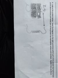
9. Look at the protein below. Label the diagram on the right and show which part of the protein has hydrophobic amino acid residues and what part of the protein has hydrophilic amino acid residues. Explain how you know.
**Diagram Explanation:**
The image shows a diagram of a protein interacting with a cell membrane. The diagram illustrates the following components:
- **Cell Membrane:**
- The membrane is depicted as a double layer of circles with tails, representing the phospholipid bilayer. The circles (heads) are polar (hydrophilic), and the tails are nonpolar (hydrophobic).
- **Protein:**
- A protein is shown weaving through the membrane, with segments inside and outside the bilayer.
- The part of the protein embedded within the membrane (across the hydrophobic tails) is typically composed of hydrophobic amino acid residues. This is because the interior of the cell membrane is hydrophobic, favoring the presence of nonpolar residues.
- The segments of the protein protruding outside the cell membrane are likely to have hydrophilic amino acid residues. These regions interact with the aqueous environment inside and outside the cell.
**Labeling and Explanation:**
- **Hydrophobic Regions:**
- These are located where the protein segments embed within the membrane, aligning with the tails of the phospholipids. This indicates nonpolar, hydrophobic residues that avoid water.
- **Hydrophilic Regions:**
- These are the portions of the protein that extend outside the membrane, interacting with the watery environment. These protein parts contain polar, hydrophilic residues that attract water.
The arrow in the diagram indicates the direction or span of the protein across the membrane, emphasizing the distinct regions based on residue interaction characteristics.
Expert Solution
This question has been solved!
Explore an expertly crafted, step-by-step solution for a thorough understanding of key concepts.
This is a popular solution
Trending nowThis is a popular solution!
Step by stepSolved in 2 steps with 1 images

Knowledge Booster
Learn more about
Need a deep-dive on the concept behind this application? Look no further. Learn more about this topic, biology and related others by exploring similar questions and additional content below.Similar questions
- 1. Theoretically, a protein could assume a virtually infinite number of configurations and conformations. Suggestseveral features of proteins that drastically limit the actual number. 2. What are the greatest structural features that differentiate sphingolipids from phosphoglycerides? 3. High levels of glucose-6-phosphate inhibit glycolysis. If the concentration of glucose-6-phosphate decreases,activity is restored. Why?arrow_forward5. Classify each of the following amino acids as polar or nonpolar and hydrophilic and hydrophobic. NH2 CH2 CH2 H2N-C-COOH H2N-C-COOH H. a. b. Polar or nonpolar: Hydrophilic or Hydrophobic:arrow_forwardOn 4. Below is a polypeptide with an unknown number of amino acids. On the diagram below: a. Circle EVERY peptide bond b. Circle and number each amino acid from 1, 2, 3, and so on; from left to right c. Label each amino acid as polar, non-polar, or electrically charged d. Label the N-terminus and C-terminus e. Select three pairs of amino acids that could potentially interact to influence tertiary structure AND state the bond/force/interaction type involved in each pair. H₂N-CH-C-N-CH-C- CH₂ CH₂ CH₂ NH CINH NH₂ Accessibility: Investigate CH₂ -NH -CH-C-N- CH-OH CH3 -CH-C-N CH₂ CH₂ -N- OH CHIC-OH CH₂ OHarrow_forward
arrow_back_ios
arrow_forward_ios
Recommended textbooks for you
 Human Anatomy & Physiology (11th Edition)BiologyISBN:9780134580999Author:Elaine N. Marieb, Katja N. HoehnPublisher:PEARSON
 Biology 2eBiologyISBN:9781947172517Author:Matthew Douglas, Jung Choi, Mary Ann ClarkPublisher:OpenStax
 Anatomy & PhysiologyBiologyISBN:9781259398629Author:McKinley, Michael P., O'loughlin, Valerie Dean, Bidle, Theresa StouterPublisher:Mcgraw Hill Education,
 Molecular Biology of the Cell (Sixth Edition)BiologyISBN:9780815344322Author:Bruce Alberts, Alexander D. Johnson, Julian Lewis, David Morgan, Martin Raff, Keith Roberts, Peter WalterPublisher:W. W. Norton & Company
 Laboratory Manual For Human Anatomy & PhysiologyBiologyISBN:9781260159363Author:Martin, Terry R., Prentice-craver, CynthiaPublisher:McGraw-Hill Publishing Co.
 Inquiry Into Life (16th Edition)BiologyISBN:9781260231700Author:Sylvia S. Mader, Michael WindelspechtPublisher:McGraw Hill Education

Human Anatomy & Physiology (11th Edition)
Biology
ISBN:9780134580999
Author:Elaine N. Marieb, Katja N. Hoehn
Publisher:PEARSON

Biology 2e
Biology
ISBN:9781947172517
Author:Matthew Douglas, Jung Choi, Mary Ann Clark
Publisher:OpenStax

Anatomy & Physiology
Biology
ISBN:9781259398629
Author:McKinley, Michael P., O'loughlin, Valerie Dean, Bidle, Theresa Stouter
Publisher:Mcgraw Hill Education,

Molecular Biology of the Cell (Sixth Edition)
Biology
ISBN:9780815344322
Author:Bruce Alberts, Alexander D. Johnson, Julian Lewis, David Morgan, Martin Raff, Keith Roberts, Peter Walter
Publisher:W. W. Norton & Company

Laboratory Manual For Human Anatomy & Physiology
Biology
ISBN:9781260159363
Author:Martin, Terry R., Prentice-craver, Cynthia
Publisher:McGraw-Hill Publishing Co.

Inquiry Into Life (16th Edition)
Biology
ISBN:9781260231700
Author:Sylvia S. Mader, Michael Windelspecht
Publisher:McGraw Hill Education