Human Anatomy & Physiology (11th Edition)
11th Edition
ISBN: 9780134580999
Author: Elaine N. Marieb, Katja N. Hoehn
Publisher: PEARSON
expand_more
expand_more
format_list_bulleted
Concept explainers
Question

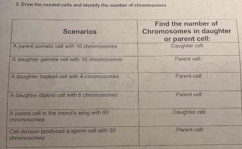
Transcribed Image Text:3. Draw the needed cells and identify the number of chromosomes
Scenarios
A parent somatic cell with 10 chromosomes
A daughter gamete cell with 10 chromosomes
A daughter haploid cell with 6 chromosomes
A daughter diploid cell with 6 chromosomes
A parent cell in the insect's wing with 80
chromosomes
Cell division produced a sperm cell with 50
chromosomes
Find the number of
Chromosomes in daughter
or parent cell:
Daughter cell:
Parent cell:
Parent cell:
Parent cell:
Daughter cell:
Parent cell:
Expert Solution
This question has been solved!
Explore an expertly crafted, step-by-step solution for a thorough understanding of key concepts.
Step by stepSolved in 2 steps with 1 images

Knowledge Booster
Learn more about
Need a deep-dive on the concept behind this application? Look no further. Learn more about this topic, biology and related others by exploring similar questions and additional content below.Similar questions
- ..explain why meiosis occurs only in specialized cells (gametes), and that the overall goal of meiosis is to make haploid cells for sexual reproduction • ..outline the sequence of key chromosomal movements and rearrangements during the two meiotic divisions, identifying key similarities and differences between meiosis and mitosis • ..describe the ploidy of a cell before and after meiosis I and meiosis II, and how ploidy changes after separation of sister chromatids and homologous chromosomesarrow_forward6)From the menu on the left on Virtual Urchin, click on "embryogenesis to hatching". This will display a video of sea urchin development. Photos of key stages taken with a scanning electron microscope (SEM) are shown next to the video. In addition to watching the video by clicking the play button, you can use the slider at the bottom to move through the stages step by step. Doing this will allow you watch how cell division increases the number of cells from the initial zygote. You'll notice that these divisions start off dividing cells evenly, but at a certain point, there are some smaller cells created. These smaller cells will play very important roles in the later development stages of the urchin, so their creation is a key milestone. Which round of cell division is uneven? (hint: the SEM images will be helpful) a)First b)Second c)Third d)Fourth e)Fiftharrow_forwardWhich of the following best describes the kinetochore? the core of proteins that forms the metaphase plate in a dividing cell a structure composed of several proteins that associate with the centromere region of a chromosome and that can bind to spindle microtubules the ring of actin microfilaments that will cause the appearance of the cleavage furrow the centromere region of a metaphase chromosome at which the DNA can bind with spindle proteinsarrow_forward
- 1. Fill in this table: Mitosis Meiosis Type of reproduction (sexual or asexual) # of cell/nuclear divisions Synapse/ crossing over? (yes or no) Centromeres dissolve Meiosis 1? during anaphase? Meiosis II? Chromosome # at beginning? Chromosome # at end of process? Meiosis I? Meiosis II? # of daughter nuclei? Daughter cells the same as parent cell? Daughter cells genetically identical to cach other? 2. a. A cell has n=2. How many total chromosomes are in a diploid cell? b. Draw this cell during metaphase of mitosis:arrow_forwardThe figure shows chromosomes, and sister chromatids in a cell. Which of the following are true? Select all that apply This is a diploid cell There are total six chromosomes present in the cell There are 12 chromatids The value ofn is 3arrow_forward2. The amount of DNA in a Drosophila melanogaster somatic cell equals 8 during G1 of interphase, how much DNA is present in the cell during each phase of mitosis and meiosis? Amount of G2 Phase Prophase Metaphase Anaphase Telophase DNA in: Mitosis 8. Meiosis I 8. Meiosis II 4arrow_forward
- mat Arrange Tools Slide Show Window Help A 12% D Sat 2:02 PM 2 Chapter 13 clicker questions imations Slide Show Review View Shape Format O Tell me e Share V Play Narrations Always Use Subtitles V Use Timings Rehearse Record Timings Slide Show V Show Media Controls A Subtitle Settings v What allows sister chromatids to finally separate, and in which phase of meiosis does this occur? a) release of cohesin along sister chromatid arms in anaphase I b) crossing over of chromatids in prophase I c) release of ohesin at centromeres in anaphase I d) release of cohesin at centromeres in anaphase II e) crossing over of homologs in prophase I O 2017 Pearson Education, Inc 103% :: E Notes Comments étv MacBook Pro 80 888 * ( & $ 8 9 6 7arrow_forwardmitosis nuclei resides in the nucleus karyotype autosomes haploid In dividing parent cell; in the parent cell. Male and female gametes at fertilization forms a homologs Microscopic observations showed that during fertilization, the unite, providing evidence that genetic material resides in the cell membrane Brightly staining, threadlike bodies within the nucleus were termed of these bodies could be traced through cell division. chromosomes diploid A image can reveal abnormalities in meiosis of eggs and sperm ; the movements gametes contain a single set of chromosomes. The fusion of zygote. the number of chromosomes in daughter cells remains the same as in the the number of chromosomes in daughter cells is half that of Two chromosomes that match in size, shape, and banding when isolated and stained are is an image of an individual's chromosomes arranged in homologous pairs. This chromosome number and structure nondisjunctionarrow_forwardWhen chromatids are separated during anaphase, how do we know that the microtubules that they are connected to are not actually shortening to "pull" the chromatids apart?arrow_forward
- Need help right awayarrow_forwardDRAW OUT CLEARLY each of these chromosomes labeling as appropriate (demoed in class) The cell has a diploid number of 8 Each pair has a set of linked genes that are located on each end of the chromosome (paternal on left, maternal on right) Pair 1 AB and ab Pair 2 DE and de Pair 3 Fg and fG Pair 4 hI and Hi Show what the chromosomes would look like in G1, G2, TI and TIIarrow_forward8arrow_forward
arrow_back_ios
SEE MORE QUESTIONS
arrow_forward_ios
Recommended textbooks for you
 Human Anatomy & Physiology (11th Edition)BiologyISBN:9780134580999Author:Elaine N. Marieb, Katja N. HoehnPublisher:PEARSON
 Biology 2eBiologyISBN:9781947172517Author:Matthew Douglas, Jung Choi, Mary Ann ClarkPublisher:OpenStax
 Anatomy & PhysiologyBiologyISBN:9781259398629Author:McKinley, Michael P., O'loughlin, Valerie Dean, Bidle, Theresa StouterPublisher:Mcgraw Hill Education,
 Molecular Biology of the Cell (Sixth Edition)BiologyISBN:9780815344322Author:Bruce Alberts, Alexander D. Johnson, Julian Lewis, David Morgan, Martin Raff, Keith Roberts, Peter WalterPublisher:W. W. Norton & Company
 Laboratory Manual For Human Anatomy & PhysiologyBiologyISBN:9781260159363Author:Martin, Terry R., Prentice-craver, CynthiaPublisher:McGraw-Hill Publishing Co.
 Inquiry Into Life (16th Edition)BiologyISBN:9781260231700Author:Sylvia S. Mader, Michael WindelspechtPublisher:McGraw Hill Education

Human Anatomy & Physiology (11th Edition)
Biology
ISBN:9780134580999
Author:Elaine N. Marieb, Katja N. Hoehn
Publisher:PEARSON

Biology 2e
Biology
ISBN:9781947172517
Author:Matthew Douglas, Jung Choi, Mary Ann Clark
Publisher:OpenStax

Anatomy & Physiology
Biology
ISBN:9781259398629
Author:McKinley, Michael P., O'loughlin, Valerie Dean, Bidle, Theresa Stouter
Publisher:Mcgraw Hill Education,

Molecular Biology of the Cell (Sixth Edition)
Biology
ISBN:9780815344322
Author:Bruce Alberts, Alexander D. Johnson, Julian Lewis, David Morgan, Martin Raff, Keith Roberts, Peter Walter
Publisher:W. W. Norton & Company

Laboratory Manual For Human Anatomy & Physiology
Biology
ISBN:9781260159363
Author:Martin, Terry R., Prentice-craver, Cynthia
Publisher:McGraw-Hill Publishing Co.

Inquiry Into Life (16th Edition)
Biology
ISBN:9781260231700
Author:Sylvia S. Mader, Michael Windelspecht
Publisher:McGraw Hill Education