Concept explainers
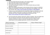
What type of symmetry is seen in the roundworm?
Does it exhibit cephalization?
Ascaris is a
View the Trichinella slide. Trichinella is also a parasite. It can infect humans as well as other mammals like pigs, bears, and rodents. If untreated, it can lead to death.
What mammal tissue does this roundworm infect?
Draw a picture below of the Trichinella as viewed under the microscope.
Although not mentioned on the website, if available view the live vinegar eel specimens. If there are no live specimens available, check out this you tube video instead: https://www.youtube.com/watch?v=UnjwvtFvyeQ
Describe the movement of the vinegar eels.
Do they have a complete or incomplete


Trending nowThis is a popular solution!
Step by stepSolved in 2 steps

- Match each structure with its descriptions. _____ radula a. internal skeleton _____ exoskeleton b. external skeleton _____ endoskeleton c. stinging cell of cnidarians _____ cnidocyte d. excretory organ of insects _____ compound eye e. mollusk feeding device _____ tube foot f. larva of annelid or mollusk _____ trochophore g. has many lenses _____ Malpighian tubule h. moves an echinoderm _____ gastrovascular cavity i. digests a planarians foodarrow_forwardThe large central opening in the parazoan body is called the: gem mule spicule ostia osculum.arrow_forwardMatch the organisms with their descriptions. _____mollusks a. complete gut, pseudocoelom _____echinoderms b. nematocyst producers _____sponges c. simplest organ systems _____cnidarians d. no tissues, filters out food _____flatworms e. jointed exoskeleton _____roundworms f. mantle over body mass _____annelids g. segmented worms _____arthropods h. tube feet, spiny skinarrow_forward
- Most sponge body plans are slight variations on a simple tube-within-a-tube design. Which of the following is a key limitation of sponge body plans? Sponges lack the specialized cell types needed to produce more complex body plans The reliance on osmosis/diffusion requires a design that maximizes the surface area to volume ratio of the sponge Choanocytes must be protected from the hostile exterior environment Spongin cannot support heavy bodies.arrow_forwardWhich group of fllatworms are primarily ectoparasites of fish? monogeneans trematodes cestodes turbellariansarrow_forwardWhich of the following features does not distinguish humans as a member of phylum Chordata? Human embryos undergo indeterminate cleavage A spinal cord runs along an adult human’s dorsal side Human embryos exhibit pharyngeal arches and gill slits, The human coccyx forms from an embryonic tail.arrow_forward
- What is the major morphological innovation seen in annelid worms? a. a complete digestive system b. image-forming eyes c. a respiratory system d. an open circulatory system e. body segmentationarrow_forwardThe rhynchocoel is a. circulatory system fluid-filled cavity primitive excretory system proboscisarrow_forwardWhich of the following is not possible? radially symmetrical diploblast diploblastic eucoelomate protostomic coelomate bilaterally symmetrical deuterostomearrow_forward
- A mantle and mantle cavity are present in: phylum Echinodermata phylum Adversoidea phylum Mollusca phylum Nemertea.arrow_forwardWhich organ system is absent in flatworms (phylum Platyhelminthes)? a. nervous system b. reproductive system c. circulatory system d. digestive system e. excretory systemarrow_forwardWhich of the following is not contained in phylum Chordata? Cephalochordata Echinodermata Urochordata Vertebrataarrow_forward
 Biology 2eBiologyISBN:9781947172517Author:Matthew Douglas, Jung Choi, Mary Ann ClarkPublisher:OpenStax
 Biology: The Unity and Diversity of Life (MindTap...BiologyISBN:9781337408332Author:Cecie Starr, Ralph Taggart, Christine Evers, Lisa StarrPublisher:Cengage Learning
 Biology: The Unity and Diversity of Life (MindTap...BiologyISBN:9781305073951Author:Cecie Starr, Ralph Taggart, Christine Evers, Lisa StarrPublisher:Cengage Learning
 Biology: The Dynamic Science (MindTap Course List)BiologyISBN:9781305389892Author:Peter J. Russell, Paul E. Hertz, Beverly McMillanPublisher:Cengage Learning
 Biology (MindTap Course List)BiologyISBN:9781337392938Author:Eldra Solomon, Charles Martin, Diana W. Martin, Linda R. BergPublisher:Cengage Learning


