Human Anatomy & Physiology (11th Edition)
11th Edition
ISBN: 9780134580999
Author: Elaine N. Marieb, Katja N. Hoehn
Publisher: PEARSON
expand_more
expand_more
format_list_bulleted
Question

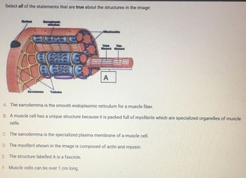
Transcribed Image Text:Select all of the statements that are true about the structures in the image:
Nucleus
Sarcoplasmic
135136
Sarcolemma T-tubules
€
Mitochondria
Thick
Blament
A
A. The sarcolemma is the smooth endoplasmic reticulum for a muscle fiber.
B. A muscle cell has a unique structure because it is packed full of myofibrils which are specialized organelles of muscle
cells.
C. The sarcolemma is the specialized plasma membrane of a muscle cell.
D. The myofibril shown in the image is composed of actin and myosin.
E. The structure labelled A is a fascicle.
F. Muscle cells can be over 1 cm long.

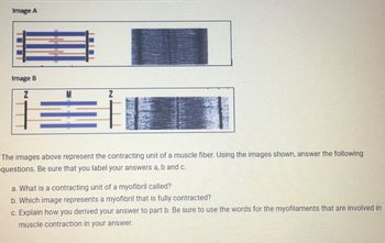
Transcribed Image Text:Image A
Image B
Z
M
F.
The images above represent the contracting unit of a muscle fiber. Using the images shown, answer the following
questions. Be sure that you label your answers a, b and c.
a. What is a contracting unit of a myofibril called?
b. Which image represents a myofibril that is fully contracted?
c. Explain how you derived your answer to part b. Be sure to use the words for the myofilaments that are involved in
muscle contraction in your answer.
Expert Solution
This question has been solved!
Explore an expertly crafted, step-by-step solution for a thorough understanding of key concepts.
This is a popular solution
Trending nowThis is a popular solution!
Step by stepSolved in 4 steps

Knowledge Booster
Similar questions
- The fold of mucus membrane containing elastic fibers that are responsible for sounds are called the ___________.arrow_forwardThe part of the brain responsible for our conscious mind white matter the cerebral cortex The thalamus the cerebellumarrow_forwardWhich dissection is best to do a biological drawing of the corpus callosumarrow_forward
- Why should your genomic make up (genes) and anatomical brain structure NOT be admissible in the courtroom? Explain why someone's genomic make up (genes) and anatomical brain structure is important information in criminal cases.arrow_forwardLabel 47,44,37arrow_forwardthe cephalic region is blank to the sternal regionarrow_forward
- True or False: The areas of cortex devoted to processing information from the fovea are larger than those devoted to processing information from the periphery, which is another reason why images projected on the fovea are seen in great detail.arrow_forwardDysfunction of which the following structures may be the cause of lower extremity symptoms? Brachial plexus Lumbar plexus Cervical plexus Sacral plexusarrow_forwardA human zygote contains ___________ chromosomes.arrow_forward
arrow_back_ios
SEE MORE QUESTIONS
arrow_forward_ios
Recommended textbooks for you
 Human Anatomy & Physiology (11th Edition)Anatomy and PhysiologyISBN:9780134580999Author:Elaine N. Marieb, Katja N. HoehnPublisher:PEARSON
 Anatomy & PhysiologyAnatomy and PhysiologyISBN:9781259398629Author:McKinley, Michael P., O'loughlin, Valerie Dean, Bidle, Theresa StouterPublisher:Mcgraw Hill Education,
 Human AnatomyAnatomy and PhysiologyISBN:9780135168059Author:Marieb, Elaine Nicpon, Brady, Patricia, Mallatt, JonPublisher:Pearson Education, Inc.,
 Anatomy & Physiology: An Integrative ApproachAnatomy and PhysiologyISBN:9780078024283Author:Michael McKinley Dr., Valerie O'Loughlin, Theresa BidlePublisher:McGraw-Hill Education
 Human Anatomy & Physiology (Marieb, Human Anatomy...Anatomy and PhysiologyISBN:9780321927040Author:Elaine N. Marieb, Katja HoehnPublisher:PEARSON

Human Anatomy & Physiology (11th Edition)
Anatomy and Physiology
ISBN:9780134580999
Author:Elaine N. Marieb, Katja N. Hoehn
Publisher:PEARSON

Anatomy & Physiology
Anatomy and Physiology
ISBN:9781259398629
Author:McKinley, Michael P., O'loughlin, Valerie Dean, Bidle, Theresa Stouter
Publisher:Mcgraw Hill Education,

Human Anatomy
Anatomy and Physiology
ISBN:9780135168059
Author:Marieb, Elaine Nicpon, Brady, Patricia, Mallatt, Jon
Publisher:Pearson Education, Inc.,

Anatomy & Physiology: An Integrative Approach
Anatomy and Physiology
ISBN:9780078024283
Author:Michael McKinley Dr., Valerie O'Loughlin, Theresa Bidle
Publisher:McGraw-Hill Education

Human Anatomy & Physiology (Marieb, Human Anatomy...
Anatomy and Physiology
ISBN:9780321927040
Author:Elaine N. Marieb, Katja Hoehn
Publisher:PEARSON