Human Anatomy & Physiology (11th Edition)
11th Edition
ISBN: 9780134580999
Author: Elaine N. Marieb, Katja N. Hoehn
Publisher: PEARSON
expand_more
expand_more
format_list_bulleted
Question
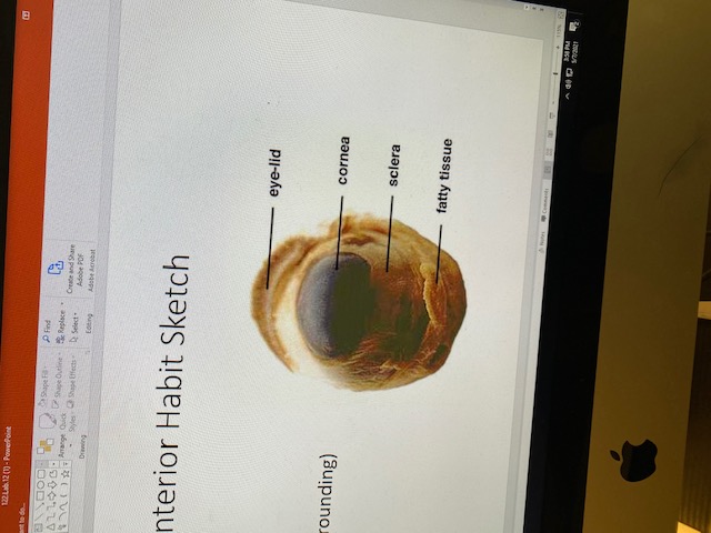
Sheep Eye
Please refer to the image to answer the question I have
Describe the texture, color, and function of the sheep eye. Does the iris change size at all (if so what causes it to change)? Is the cornea clear or cloudy?

Expert Solution
This question has been solved!
Explore an expertly crafted, step-by-step solution for a thorough understanding of key concepts.
Step by stepSolved in 2 steps

Knowledge Booster
Learn more about
Need a deep-dive on the concept behind this application? Look no further. Learn more about this topic, biology and related others by exploring similar questions and additional content below.Similar questions
- If you look at 20/20 line and it was in focus where in the eye was the image projected onto of a person's eyesight is worse than 20/20, where is that image projected?arrow_forwardI am needing clarification of why cochlear implants bypass damaged hair cells?arrow_forwardSheep Eye Please refer to the image to answer the question I have Explain the color, textures, and functions of the parts of the eye. Explain any diseases and other conditions of the eye (cataracts, detached retina, macular degeneration, near-and farsightedness) and how they relate to the structure of the eye? What new laser-based surgeries are used to correct vision? Do they act on the lens, the cornea, or the retina?arrow_forward
- Please label the following structure: A. Retina B. Sclera C. Choroid D. Corneaarrow_forwardretina disease in diabetes and hypertension Importance of prevention (hint: regular retina exam with ophthalmoscope: what are signs of concern)arrow_forwardSelect all structures that are part of the outermost layer of the eye. Sclera Cornea Uvea Choroid Retinaarrow_forward
- Recall that the eye is composed of three layers or "tunics" — the fibrous, vascular, and nervous layers — which enclose two cavities that are separated from each other by the lens. Review the components of these three layers by matching each description with the appropriate letter in the figure below: 1. Fluid in the anterior portion of the eye that provides nutrients to the lens and cornea 2. The "whites" of the eye 3. Area of the retina that lacks photoreceptors 4. Contains smooth muscle that controls the shape of the lens 5. Nutritive (nourishing) layer of the eye 6. Layer containing rods and cones 7. Gel-like substance that helps support the eyeball 8. Pigmented smooth muscles that control pupil size 9. Most anterior component of the fibrous layer — your "window to the world" 10. Structure that changes shape to bend light toward rods and conesarrow_forwardThe Ishihara test is used to test for color blindness visual acuity astigmatism depth perceptionarrow_forwardGive typed explanation of all subparts otherwise leave it Within the textbox below, define and identify the combining forms (Root, Suffix, Prefix, Combining vowel) of the following terms: Example: Salpingopharyngeal = salping/o-pharyng-eal 4. Otoscope= 5. Rhinitis 6. Amblyopia 7. Astigmatism= 8. Corneal 9. Xerosis 10. Audiogramarrow_forward
- Presbyopia is a condition in which a person's ability to adjust to changes in visual acuity diminishes or disappears entirely. Will she still require reading glasses after LASIK to correct her far vision? Explain.arrow_forward(a) What is hypermetropia?(b) What are the two causes of this defect of vision?arrow_forwardIf I had a eyesight that was measured as being 20/10 _________. What would happen .arrow_forward
arrow_back_ios
SEE MORE QUESTIONS
arrow_forward_ios
Recommended textbooks for you
 Human Anatomy & Physiology (11th Edition)BiologyISBN:9780134580999Author:Elaine N. Marieb, Katja N. HoehnPublisher:PEARSON
 Biology 2eBiologyISBN:9781947172517Author:Matthew Douglas, Jung Choi, Mary Ann ClarkPublisher:OpenStax
 Anatomy & PhysiologyBiologyISBN:9781259398629Author:McKinley, Michael P., O'loughlin, Valerie Dean, Bidle, Theresa StouterPublisher:Mcgraw Hill Education,
 Molecular Biology of the Cell (Sixth Edition)BiologyISBN:9780815344322Author:Bruce Alberts, Alexander D. Johnson, Julian Lewis, David Morgan, Martin Raff, Keith Roberts, Peter WalterPublisher:W. W. Norton & Company
 Laboratory Manual For Human Anatomy & PhysiologyBiologyISBN:9781260159363Author:Martin, Terry R., Prentice-craver, CynthiaPublisher:McGraw-Hill Publishing Co.
 Inquiry Into Life (16th Edition)BiologyISBN:9781260231700Author:Sylvia S. Mader, Michael WindelspechtPublisher:McGraw Hill Education

Human Anatomy & Physiology (11th Edition)
Biology
ISBN:9780134580999
Author:Elaine N. Marieb, Katja N. Hoehn
Publisher:PEARSON

Biology 2e
Biology
ISBN:9781947172517
Author:Matthew Douglas, Jung Choi, Mary Ann Clark
Publisher:OpenStax

Anatomy & Physiology
Biology
ISBN:9781259398629
Author:McKinley, Michael P., O'loughlin, Valerie Dean, Bidle, Theresa Stouter
Publisher:Mcgraw Hill Education,

Molecular Biology of the Cell (Sixth Edition)
Biology
ISBN:9780815344322
Author:Bruce Alberts, Alexander D. Johnson, Julian Lewis, David Morgan, Martin Raff, Keith Roberts, Peter Walter
Publisher:W. W. Norton & Company

Laboratory Manual For Human Anatomy & Physiology
Biology
ISBN:9781260159363
Author:Martin, Terry R., Prentice-craver, Cynthia
Publisher:McGraw-Hill Publishing Co.

Inquiry Into Life (16th Edition)
Biology
ISBN:9781260231700
Author:Sylvia S. Mader, Michael Windelspecht
Publisher:McGraw Hill Education