Human Anatomy & Physiology (11th Edition)
11th Edition
ISBN: 9780134580999
Author: Elaine N. Marieb, Katja N. Hoehn
Publisher: PEARSON
expand_more
expand_more
format_list_bulleted
Question
Please color it

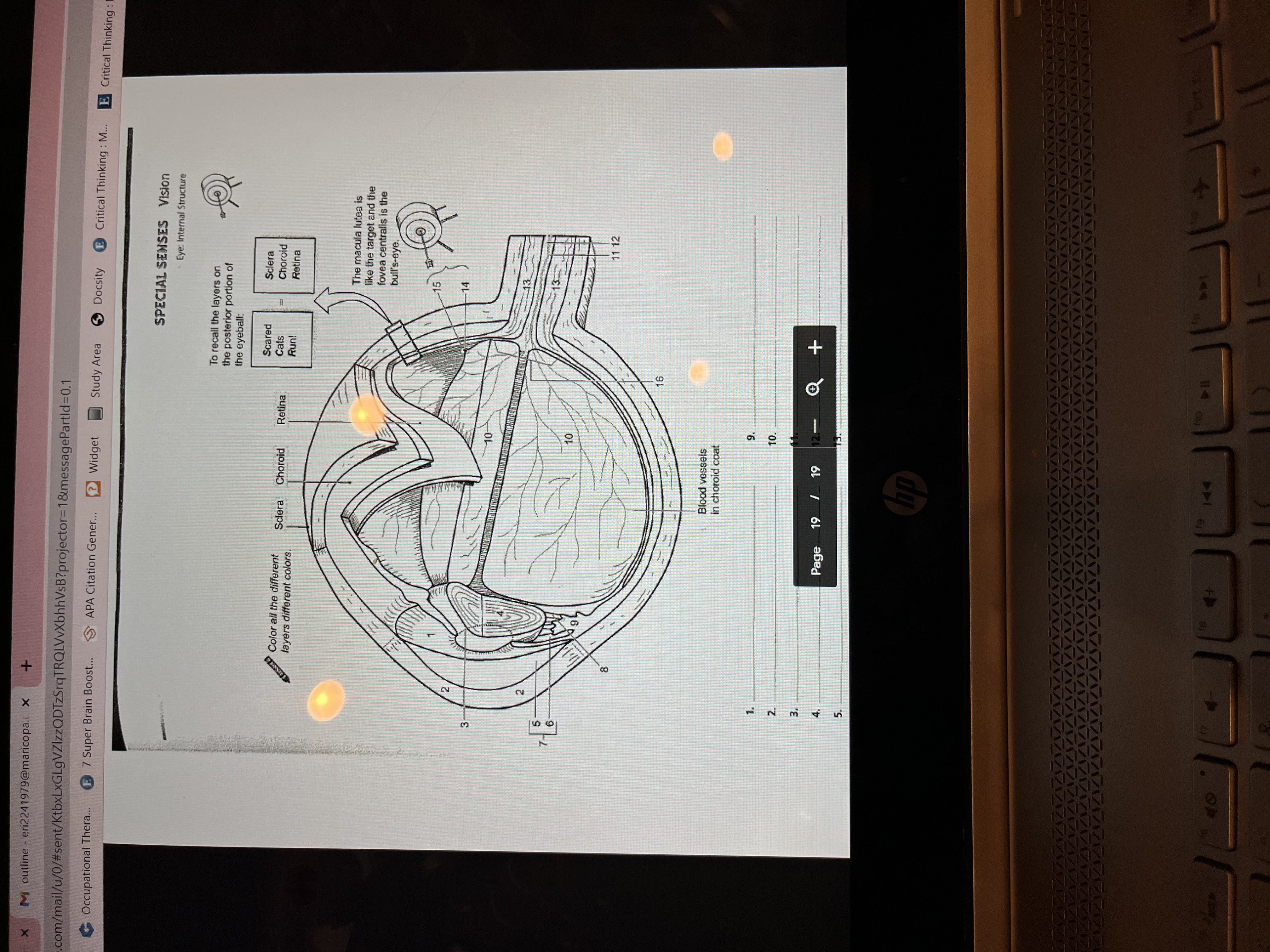
Transcribed Image Text:**Special Senses: Vision**
**Eye: Internal Structure**
This educational diagram provides a detailed view of the internal structure of the eye, highlighting the different layers and components. Each part is numbered for reference.
### Diagram Description
1. **Sclera**: The outer layer, forming the white of the eye.
2. **Choroid**: The middle layer, rich in blood vessels.
3. **Retina**: The innermost layer containing light-sensitive cells.
### Key Features:
- **Color Coding Task**: It suggests coloring each layer (sclera, choroid, retina) a different color for better understanding.
- **Mnemonic Device**: "Scared Cats Run" helps remember the order of the layers: Sclera, Choroid, Retina.
### Additional Annotations:
- **Macula Lutea**: Highlighted as the target, with the fovea centralis as the bull's-eye.
- **Blood Vessels**: Shown in the choroid coat, essential for supplying nutrients to the eye.
#### Notations on Diagram:
1. (empty)
2. (empty)
3. (empty)
4. (empty)
5. (empty)
6. (empty)
7. (empty)
8. (empty)
9. (empty)
10. (empty)
11. (empty)
12. (empty)
13. (empty)
14. (empty)
15. (empty)
16. (with note: blood vessels in choroid coat)
This diagram serves as a study aid for understanding the complex anatomy of the eye and the function of its different layers.
Expert Solution
This question has been solved!
Explore an expertly crafted, step-by-step solution for a thorough understanding of key concepts.
This is a popular solution
Trending nowThis is a popular solution!
Step by stepSolved in 2 steps with 1 images

Knowledge Booster
Similar questions
- Can you help me to write the name of the label of this picture? I have a hard time to figure it out.arrow_forwardExplain the additional health risks to a patient undergoing a CT scan as compared to an ordinary X-ray photograph. 150 words. Please could you write it on paper.arrow_forwardCan you help me the name of the labels?arrow_forward
arrow_back_ios
arrow_forward_ios
Recommended textbooks for you
 Human Anatomy & Physiology (11th Edition)Anatomy and PhysiologyISBN:9780134580999Author:Elaine N. Marieb, Katja N. HoehnPublisher:PEARSON
 Anatomy & PhysiologyAnatomy and PhysiologyISBN:9781259398629Author:McKinley, Michael P., O'loughlin, Valerie Dean, Bidle, Theresa StouterPublisher:Mcgraw Hill Education,
 Human AnatomyAnatomy and PhysiologyISBN:9780135168059Author:Marieb, Elaine Nicpon, Brady, Patricia, Mallatt, JonPublisher:Pearson Education, Inc.,
 Anatomy & Physiology: An Integrative ApproachAnatomy and PhysiologyISBN:9780078024283Author:Michael McKinley Dr., Valerie O'Loughlin, Theresa BidlePublisher:McGraw-Hill Education
 Human Anatomy & Physiology (Marieb, Human Anatomy...Anatomy and PhysiologyISBN:9780321927040Author:Elaine N. Marieb, Katja HoehnPublisher:PEARSON

Human Anatomy & Physiology (11th Edition)
Anatomy and Physiology
ISBN:9780134580999
Author:Elaine N. Marieb, Katja N. Hoehn
Publisher:PEARSON

Anatomy & Physiology
Anatomy and Physiology
ISBN:9781259398629
Author:McKinley, Michael P., O'loughlin, Valerie Dean, Bidle, Theresa Stouter
Publisher:Mcgraw Hill Education,

Human Anatomy
Anatomy and Physiology
ISBN:9780135168059
Author:Marieb, Elaine Nicpon, Brady, Patricia, Mallatt, Jon
Publisher:Pearson Education, Inc.,

Anatomy & Physiology: An Integrative Approach
Anatomy and Physiology
ISBN:9780078024283
Author:Michael McKinley Dr., Valerie O'Loughlin, Theresa Bidle
Publisher:McGraw-Hill Education

Human Anatomy & Physiology (Marieb, Human Anatomy...
Anatomy and Physiology
ISBN:9780321927040
Author:Elaine N. Marieb, Katja Hoehn
Publisher:PEARSON