Human Anatomy & Physiology (11th Edition)
11th Edition
ISBN: 9780134580999
Author: Elaine N. Marieb, Katja N. Hoehn
Publisher: PEARSON
expand_more
expand_more
format_list_bulleted
Question
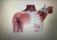
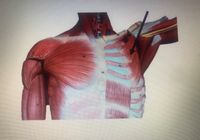
Label the structure
A. Pectoralis minor
B. Pectoralis major
C. Trapezius
D. Latissimus dorsi
E. Teres minor

Transcribed Image Text:The image shows a detailed anatomical illustration of the upper torso, highlighting the musculature around the shoulder region. The focus is on the pectoralis minor muscle, as indicated by the black arrow. The pectoralis minor is located beneath the larger pectoralis major muscle and is one of the key muscles involved in the movement of the scapula.
The muscles are shown in a realistic reddish hue, with fibers and contours illustrating their structure and direction. The diagram indicates surrounding areas, including part of the rib cage to which the pectoralis minor attaches. Proper understanding of this muscle's position and function is essential for fields such as medicine, physiotherapy, and sports science.
* A. Pectoralis minor
This depiction serves as an educational aid in understanding the anatomical relationships and functions of the muscles in the shoulder and chest region.

Transcribed Image Text:The image depicts the muscles of the upper torso, particularly focusing on the pectoral region. The major muscle groups visible include:
1. **Pectoralis Major**: This large muscle covers the upper chest and contributes to the movement of the shoulder joint. It is shown prominently across the chest area.
2. **Serratus Anterior**: Located on the lateral aspect of the thorax, these muscles appear serrated and are involved in the upward rotation and protraction of the scapula.
3. **Intercostal Muscles**: These muscles are located between the ribs and are involved in the mechanical aspect of breathing by aiding in the expansion and contraction of the chest cavity.
The image also includes an arrow pointing to a specific muscle group, possibly indicating the highlighted region for educational focus. This could refer to an illustrative emphasis on the **Pectoralis Minor**, a smaller muscle situated underneath the Pectoralis Major and involved in stabilization, depression, abduction, or protraction of the scapula.
This anatomical illustration is valuable for understanding the muscular structure and arrangement in the pectoral region, aiding in the study of kinesiology and anatomy.
Expert Solution
This question has been solved!
Explore an expertly crafted, step-by-step solution for a thorough understanding of key concepts.
Step by stepSolved in 4 steps

Knowledge Booster
Similar questions
- This muscle is a pyramid shaped muscle of facial expression. This muscle lies beneath the frontalis and orbicularis oculi. a. Procerus b. Corrugator c. Risorius d. Digastricarrow_forwardWhich muscles attach to the ischial tuberosity and extend thethigh plus flex the leg?a. adductor musclesb. fibularis musclesc. hamstring musclesd. quadriceps musclesarrow_forwardThe muscle in the upper arm that is used as an injection site is thea. triceps.b. biceps.c. trapezius.d. deltoid.arrow_forward
Recommended textbooks for you
 Human Anatomy & Physiology (11th Edition)Anatomy and PhysiologyISBN:9780134580999Author:Elaine N. Marieb, Katja N. HoehnPublisher:PEARSON
 Anatomy & PhysiologyAnatomy and PhysiologyISBN:9781259398629Author:McKinley, Michael P., O'loughlin, Valerie Dean, Bidle, Theresa StouterPublisher:Mcgraw Hill Education,
 Human AnatomyAnatomy and PhysiologyISBN:9780135168059Author:Marieb, Elaine Nicpon, Brady, Patricia, Mallatt, JonPublisher:Pearson Education, Inc.,
 Anatomy & Physiology: An Integrative ApproachAnatomy and PhysiologyISBN:9780078024283Author:Michael McKinley Dr., Valerie O'Loughlin, Theresa BidlePublisher:McGraw-Hill Education
 Human Anatomy & Physiology (Marieb, Human Anatomy...Anatomy and PhysiologyISBN:9780321927040Author:Elaine N. Marieb, Katja HoehnPublisher:PEARSON

Human Anatomy & Physiology (11th Edition)
Anatomy and Physiology
ISBN:9780134580999
Author:Elaine N. Marieb, Katja N. Hoehn
Publisher:PEARSON

Anatomy & Physiology
Anatomy and Physiology
ISBN:9781259398629
Author:McKinley, Michael P., O'loughlin, Valerie Dean, Bidle, Theresa Stouter
Publisher:Mcgraw Hill Education,

Human Anatomy
Anatomy and Physiology
ISBN:9780135168059
Author:Marieb, Elaine Nicpon, Brady, Patricia, Mallatt, Jon
Publisher:Pearson Education, Inc.,

Anatomy & Physiology: An Integrative Approach
Anatomy and Physiology
ISBN:9780078024283
Author:Michael McKinley Dr., Valerie O'Loughlin, Theresa Bidle
Publisher:McGraw-Hill Education

Human Anatomy & Physiology (Marieb, Human Anatomy...
Anatomy and Physiology
ISBN:9780321927040
Author:Elaine N. Marieb, Katja Hoehn
Publisher:PEARSON