Human Anatomy & Physiology (11th Edition)
11th Edition
ISBN: 9780134580999
Author: Elaine N. Marieb, Katja N. Hoehn
Publisher: PEARSON
expand_more
expand_more
format_list_bulleted
Question
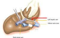
The unlabeled structure (marked by the red arrow), would in fetal life
(A) transport deoxygenated blood from the liver tissue to the inferior vena cava
(B) transport nutrient-rich, oxygenated blood from the placenta to the inferior vena cava
(C) transport nutrient-rich, oxygenated blood from the liver tissue to ther inferior vena cava
(D) transport deoxygenated blood from the placenta to the inferior vena cava

Transcribed Image Text:The image depicts a detailed anatomical illustration of the liver with emphasis on certain veins. It highlights the following components:
1. **Right Portal Vein**: Positioned towards the left side of the image, the right portal vein is shown in the context of the liver's vascular structure.
2. **Left Hepatic Vein**: Indicated with a red arrow, this vein is a critical vessel responsible for draining deoxygenated blood from the liver to the heart.
3. **Inferior Vena Cava**: Represented in the image as a large blue structure. It is one of the body's largest veins, carrying deoxygenated blood from the lower half of the body back to the heart.
The illustration seems to display a surgical or anatomical examination setting, where certain vessels may be manipulated or highlighted for educational purposes. The hand in the image suggests a focus on understanding the spatial relations of these vessels during procedures involving the liver.
Expert Solution
This question has been solved!
Explore an expertly crafted, step-by-step solution for a thorough understanding of key concepts.
This is a popular solution
Trending nowThis is a popular solution!
Step by stepSolved in 3 steps

Knowledge Booster
Similar questions
- The circulatory system of the vertebrates is derived from ? of the Cnideria: a) ectoderm, b) the endoderm, c) heart, d) chloroplasts, e) the amoebocytes which occupy the space between the ectoderm and the endoderm.arrow_forwardWhat is primary oocytes and secondary oocytes?arrow_forwardWhich of the following is the cause of this type of anemia (A) lack of iron (B) lack of Vitamin B12 (C) lack of intrinsic factor (D) single amino acid substitution (glutamic acid replaces valine) (E) single amino acid substitution (valine replaces glutamic acid) (F) loss of bone marrow stem cellsarrow_forward
- When a ______ cell encounter a foreign bacterium, it undergoes mitosis to form a _______ cell fill in blank (A) B,T (B) B, plasma (C) T, Reed- Sternberg cell (D) plasma, thrombocytearrow_forwardConcerning the mixture ofarterial with venous bloodwhat is the differencebetween the human fetalcirculation and the adultcirculation?arrow_forwardThe vessel marked in the image below (A) is a branch of the celiac trunk (B) supplies oxygenated blood to the transverse colon (C) is found between two layers of the peritoneum (D) A and B (E) A and C (F) B and C (G) all of the abovearrow_forward
arrow_back_ios
arrow_forward_ios
Recommended textbooks for you
 Human Anatomy & Physiology (11th Edition)Anatomy and PhysiologyISBN:9780134580999Author:Elaine N. Marieb, Katja N. HoehnPublisher:PEARSON
 Anatomy & PhysiologyAnatomy and PhysiologyISBN:9781259398629Author:McKinley, Michael P., O'loughlin, Valerie Dean, Bidle, Theresa StouterPublisher:Mcgraw Hill Education,
 Human AnatomyAnatomy and PhysiologyISBN:9780135168059Author:Marieb, Elaine Nicpon, Brady, Patricia, Mallatt, JonPublisher:Pearson Education, Inc.,
 Anatomy & Physiology: An Integrative ApproachAnatomy and PhysiologyISBN:9780078024283Author:Michael McKinley Dr., Valerie O'Loughlin, Theresa BidlePublisher:McGraw-Hill Education
 Human Anatomy & Physiology (Marieb, Human Anatomy...Anatomy and PhysiologyISBN:9780321927040Author:Elaine N. Marieb, Katja HoehnPublisher:PEARSON

Human Anatomy & Physiology (11th Edition)
Anatomy and Physiology
ISBN:9780134580999
Author:Elaine N. Marieb, Katja N. Hoehn
Publisher:PEARSON

Anatomy & Physiology
Anatomy and Physiology
ISBN:9781259398629
Author:McKinley, Michael P., O'loughlin, Valerie Dean, Bidle, Theresa Stouter
Publisher:Mcgraw Hill Education,

Human Anatomy
Anatomy and Physiology
ISBN:9780135168059
Author:Marieb, Elaine Nicpon, Brady, Patricia, Mallatt, Jon
Publisher:Pearson Education, Inc.,

Anatomy & Physiology: An Integrative Approach
Anatomy and Physiology
ISBN:9780078024283
Author:Michael McKinley Dr., Valerie O'Loughlin, Theresa Bidle
Publisher:McGraw-Hill Education

Human Anatomy & Physiology (Marieb, Human Anatomy...
Anatomy and Physiology
ISBN:9780321927040
Author:Elaine N. Marieb, Katja Hoehn
Publisher:PEARSON