
Human Heredity: Principles and Issues (MindTap Course List)
11th Edition
ISBN: 9781305251052
Author: Michael Cummings
Publisher: Cengage Learning
expand_more
expand_more
format_list_bulleted
Related questions
Question
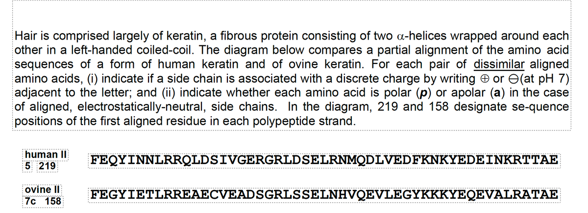

Transcribed Image Text:Hair is comprised largely of keratin, a fibrous protein consisting of two α-helices wrapped around each other in a left-handed coiled-coil. The diagram below compares a partial alignment of the amino acid sequences of a form of human keratin and of ovine keratin. For each pair of dissimilar aligned amino acids, (i) indicate if a side chain is associated with a discrete charge by writing ⊕ or ⊖ (at pH 7) adjacent to the letter; and (ii) indicate whether each amino acid is polar (p) or apolar (a) in the case of aligned, electrostatically-neutral, side chains. In the diagram, 219 and 158 designate sequence positions of the first aligned residue in each polypeptide strand.
Diagram:
- **Human Sequence (5 to 219):**
```
FEQYINNLRRQLDSIVGERGRLDSSELRNMQDLVEDFKNKYEDENKRTTAE
```
- **Ovine Sequence (7c to 158):**
```
FEGYIETLRREAECVEADSGRLSSSELNHVQEVLEGYKKYKYEQEVALRATAE
```
This alignment compares specific sequences of human and ovine keratin, focusing on differences in amino acids and their properties.
Expert Solution
This question has been solved!
Explore an expertly crafted, step-by-step solution for a thorough understanding of key concepts.

This is a popular solution

Trending nowThis is a popular solution!

Step by stepSolved in 2 steps with 1 images

Knowledge Booster

Similar questions
- Below is the structure of glycine. Draw a tripeptide composed exclusively of glycine. Label the N-terminus and C-terminus. Draw a box around the peptide bonds.arrow_forwardPolypeptide folding is often mediated by other proteins called chaperones. Describe how a mutant chaperone protein might be responsible for a genetic disorder involving an enzyme.arrow_forwardAmino acids have the generic structure seen below, where R represents different carbon-based side chains. Describe how the structure of amino acids allows them to be linked into long peptide chains to form proteins.arrow_forward
- a. Can a mutation change a proteins tertiary structure without changing its primary structure? b. Can a mutation change a proteins primary structure without affecting its secondary structure?arrow_forwardDescribe the differences in the four protein structures.arrow_forwardA beginning genetics student is attempting to complete an assignment to draw a base pair from a DNA molecule. The drawing is incomplete, and the student does not know how to finish. He asks for your advice. The assignment sheet shows that the drawing is to contain three hydrogen bonds, a purine, and a pyrimidine. From your knowledge of the pairing rules and the number of hydrogen bonds in A/T and G/C base pairs, what base pair do you help the student draw?arrow_forward
- If phenylalanine was not an essential amino acid, would diet therapy (the elimination of phenylalanine from the diet) for PKU work?arrow_forwardUnderstanding the Relevance of Chaperones in Protein Folding Protein molecules, like all molecules, can be characterized in terms of general properties such as size, shape, charge, solubility/hydrophobicity. Consider the influence of each of these general features on the likelihood of whether folding of a particular protein will require chaperone assistance or not. Be specific regarding just Hsp7O chaperones or Hsp7O chaperones and Hsp60 chaperonins.arrow_forwardWhat are Okazaki fragments and how they are formed?arrow_forward
- Which of the following levels of protein structure may be affected by hydrogen bonding? (a) primary and secondary (b) primary and tertiary (c) secondary, tertiary, and quaternary (d) primary, secondary, and tertiary (e) primary, secondary, tertiary, and quaternaryarrow_forwardUnderstanding the Nature of the Ubiquitin-Ubiquitin linkage Draw the structure of the isopeptide bond formed between Gly76of one ubiquitin molecule and Lys48of another ubiquitin molecule.arrow_forwardBelow is a sequence of 540 bases from a genome. What information would you use to find the beginnings and ends of open reading frames? How many open reading frames can you find in this sequence? Which open reading frame is likely to represent a protein- coding sequence, and why? Which are probably not functioning protein-coding sequences, and why? Note: for simplicitys sake, analyze only this one strand of the DNA double helix, reading from left to right, so you will only be analyzing three of the six reading frames shown in Figure 19.4.arrow_forward
arrow_back_ios
SEE MORE QUESTIONS
arrow_forward_ios
Recommended textbooks for you
- Human Heredity: Principles and Issues (MindTap Co...BiologyISBN:9781305251052Author:Michael CummingsPublisher:Cengage Learning

 Biology (MindTap Course List)BiologyISBN:9781337392938Author:Eldra Solomon, Charles Martin, Diana W. Martin, Linda R. BergPublisher:Cengage Learning
 Principles Of Radiographic Imaging: An Art And A ...Health & NutritionISBN:9781337711067Author:Richard R. Carlton, Arlene M. Adler, Vesna BalacPublisher:Cengage Learning
 Biology 2eBiologyISBN:9781947172517Author:Matthew Douglas, Jung Choi, Mary Ann ClarkPublisher:OpenStax
Human Heredity: Principles and Issues (MindTap Co...
Biology
ISBN:9781305251052
Author:Michael Cummings
Publisher:Cengage Learning
Biology (MindTap Course List)
Biology
ISBN:9781337392938
Author:Eldra Solomon, Charles Martin, Diana W. Martin, Linda R. Berg
Publisher:Cengage Learning
Principles Of Radiographic Imaging: An Art And A ...
Health & Nutrition
ISBN:9781337711067
Author:Richard R. Carlton, Arlene M. Adler, Vesna Balac
Publisher:Cengage Learning
Biology 2e
Biology
ISBN:9781947172517
Author:Matthew Douglas, Jung Choi, Mary Ann Clark
Publisher:OpenStax