Human Anatomy & Physiology (11th Edition)
11th Edition
ISBN: 9780134580999
Author: Elaine N. Marieb, Katja N. Hoehn
Publisher: PEARSON
expand_more
expand_more
format_list_bulleted
Question

Transcribed Image Text:**Functional Classification**
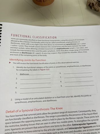
Joints are commonly classified by their functional characteristics, using the amount of movement allowed as the criterion. Functionally, joints may be called synarthroses, amphiarthroses, or diarthroses. Joints with a very tight union that allows no movement are **synarthroses** (_syn_ = union + _arthro_ = joint). They include sutures between the cranial bones and the joints between the teeth and their sockets. Joints allowing a limited movement are **amphiarthroses** (_amphi_ = two sides), and include the intervertebral discs and the pubic symphysis. Joints that allow a large range of movement are known as **diarthroses** (_di_ = apart). As you may suspect, they include the shoulder, elbow, knee, and most of the remaining joints of the body.
**Identifying Joints by Function**
1. You will review the functional classification of joints in this observational exercise.
- Identify the functional category of the joints as synarthroses, amphiarthroses, or diarthroses by completing the labels in **Figure 12.3.**
```
1. diarthroses
2. ___________
3. ___________
4. ___________
5. ___________
6. ___________
7. ___________
8. ___________
9. ___________
```
2. Using a model of an articulated skeleton or a chart from your lab, identify the joints as synarthroses, amphiarthroses, and diarthroses.
**Detail of a Synovial Diarthrosis: The Knee**
You have learned that synovial joints allow the greatest range of movement. Consequently, they are functionally classified as diarthroses. The range is provided by the presence of the liquid-filled space between the opposing bones, which is held in place by the fibrous capsule. These joints lack the restrictive, binding tissues that are seen in other less mobile joints. To strengthen the synovial joint, ligaments are present that extend from one bone to the other. **Extracapsular ligaments** are located external to the articular capsule, and **intracapsular ligaments** are found within the capsule in joints such as the knee and shoulder, one or more expansion is often seen,

Transcribed Image Text:**Patellar Ligament and Knee Joint**
The knee includes the **patellar ligament**, which is an extension of the large patellar tendon from the quadriceps muscle group of the thigh. It connects the patella to the tibial tuberosity and stabilizes the knee anteriorly. The movements of the knee joint are cushioned by numerous bursae, including the suprapatellar bursa, the subcutaneous prepatellar bursa, the deep infrapatellar bursa, and ten others.
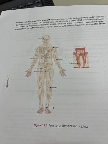
**Diagram Explanation: Functional Classification of Joints**
*Figure 12.3* presents a diagram detailing the functional classification of joints within the human body. The diagram includes:
1. **Skeletal System Illustration**: A full-body diagram highlighting key joints with labeled points:
- **Point 1**: Shoulder joint
- **Point 2**: Elbow joint
- **Point 3**: Knee joint
- **Point 4**: Ankle joint
- **Point 5**: Jaw joint (temporomandibular joint)
- **Point 6**: Hip joint
- **Point 7**: Wrist joint
- **Point 8**: Sacroiliac joint
2. **Cross-Section of a Tooth Diagram**: Displays internal components such as:
- Tooth nerve
- Tooth root
- Crown
This educational diagram is intended to enhance understanding of joint classification and anatomy, illustrating where major joints are located in the body.
*(Diagram Source: © Bluedoor, LLC)*
Expert Solution
This question has been solved!
Explore an expertly crafted, step-by-step solution for a thorough understanding of key concepts.
This is a popular solution
Trending nowThis is a popular solution!
Step by stepSolved in 3 steps

Knowledge Booster
Similar questions
- TABLE 9.1 Structural and Functional Classification of Joints Functional Classification and Amount of Motion Allowed Joint Structural Classification Structural Subcategory ual/o Intervertebral joint tion ( motor Shoulder (glenohumeral) joint lchol ole。 Intercarpal joint d ho ar jur Coronal suture ofilai Costochondral joint lame Atlantoaxial joint Tooth in its alveolus ss-b Interphalangeal joint an e 234 Exploring Anatomy & Physiology in the Laboratoryarrow_forward9.1 Structural and Functional Classification of Joints TABLE Functional Classification and Amount of Motion Allowed Structural Subcategory Structural Classification Joint hual/o Intervertebral joint tion ( motor Shoulder yIchol (glenohumeral) joint ole of Intercarpal joint nd ho lar jur Coronal suture ofilai Costochondral joint lame Atlantoaxial joint Tooth in its alveolus Interphalangeal joint ss-b an e 234 Exploring Anatomy & Physiology in the Laboratoryarrow_forwardStructural Description of Location of Joint Bone Articulations Motion Muscle(s) responsible Classification movement muscles A1 Flexion Atlas: superior Neck: Atlanto- articular facets Condylar A2 Extension Sternocleidomastoid occipital Occipital bone: occipital cond АЗ Hyperextension Neck: В 1 Atlantoaxial C1 Spine: Erector spinae group Vertebral C2 intervertebralarrow_forward
- Table 9.2 Classification of synovial jointsarrow_forwardTopic: Joints Synovial Joints are classified according to six different shapes. First, describe each type of joint shape, and then give example of where it can be found in the body.arrow_forwardThe pectoralis major can do which of the following movements at the shoulder? Group of answer choices External rotation Horizontal adduction All of these Horizontal abductionarrow_forward
- When evaluating a patient with a complaint of "twisting" the knee while playing sports. What musculoskeletal maneuver would best evaluate any type of injury and why. What is a positive vs negative exam.arrow_forwardWhat joints, in particular, occur in these planes and axis' during these ranges of motion? Shoulder Flexion (plane: sagittal, axis: frontal) Shoulder Extension (plane: sagittal, axis: frontal) Shoulder abduction (plane: frontal, axis: sagittal) Shoulder Horizontal Abduction (plane: transverse, axis: vertical)arrow_forwardA score of 1 on the FMS shoulder mobility pattern is most likely not attributed to: Joint laxity Tightness of the latissimus Doris Rounded shoulders Overdevelopment of the pectoralis minorarrow_forward
- Procedure 1 ldentifying Joint Motions of Common Movements Team up with a partner, and have your partner perform each of the following a actions are performed, and list the joints that are in motion ctions. Watch carefully as the for each joint (e.g., anual/ Ask your instructor if he or she wants you to use the technical name or the common name ious. Others, such be obv glenohumeral versus shoulder joint). Some of the joints, such as the hip joint and knee joint, will as the radio ulnar joint, the interphala ngeal joints, and the intervertebral joints, are less obvious and easily overlooked. ised, determine which motions are occurring at each joint. Keep in mind the leted the activity, answer ction Once you have listed the joints being motions are type and the range of motion of each joint as you answer each question. When you have comp Check Your Understanding question 7 (p. 252). moto 1 Walking up stairs lcho Joints moving: Motions occurring: le o dho r jui lar 2 Doing jumping jacks…arrow_forwardTree pose: Hip: Knee: Shoulder: Lunge: Hip: extenspon Knee: abduction Shoulder: With your partner, perform these common actions and determine which joints are moving and what movements are occurri Walking: Joints moving Movements occurring:arrow_forwardThe inferior tibiofibular joint's structural SUBTYPE is ______. synchondrosis condylar syndesmosis symphysis The intervertebral joint that is between the articular processes of the vertebral bones is classified functionally as _____ and structural SUBTYPE as _____. diarthrotic; symphysis diarthrotic; plane amphiarthrotic; plane amphiarthrotic; symphysisarrow_forward
arrow_back_ios
SEE MORE QUESTIONS
arrow_forward_ios
Recommended textbooks for you
 Human Anatomy & Physiology (11th Edition)Anatomy and PhysiologyISBN:9780134580999Author:Elaine N. Marieb, Katja N. HoehnPublisher:PEARSON
 Anatomy & PhysiologyAnatomy and PhysiologyISBN:9781259398629Author:McKinley, Michael P., O'loughlin, Valerie Dean, Bidle, Theresa StouterPublisher:Mcgraw Hill Education,
 Human AnatomyAnatomy and PhysiologyISBN:9780135168059Author:Marieb, Elaine Nicpon, Brady, Patricia, Mallatt, JonPublisher:Pearson Education, Inc.,
 Anatomy & Physiology: An Integrative ApproachAnatomy and PhysiologyISBN:9780078024283Author:Michael McKinley Dr., Valerie O'Loughlin, Theresa BidlePublisher:McGraw-Hill Education
 Human Anatomy & Physiology (Marieb, Human Anatomy...Anatomy and PhysiologyISBN:9780321927040Author:Elaine N. Marieb, Katja HoehnPublisher:PEARSON

Human Anatomy & Physiology (11th Edition)
Anatomy and Physiology
ISBN:9780134580999
Author:Elaine N. Marieb, Katja N. Hoehn
Publisher:PEARSON

Anatomy & Physiology
Anatomy and Physiology
ISBN:9781259398629
Author:McKinley, Michael P., O'loughlin, Valerie Dean, Bidle, Theresa Stouter
Publisher:Mcgraw Hill Education,

Human Anatomy
Anatomy and Physiology
ISBN:9780135168059
Author:Marieb, Elaine Nicpon, Brady, Patricia, Mallatt, Jon
Publisher:Pearson Education, Inc.,

Anatomy & Physiology: An Integrative Approach
Anatomy and Physiology
ISBN:9780078024283
Author:Michael McKinley Dr., Valerie O'Loughlin, Theresa Bidle
Publisher:McGraw-Hill Education

Human Anatomy & Physiology (Marieb, Human Anatomy...
Anatomy and Physiology
ISBN:9780321927040
Author:Elaine N. Marieb, Katja Hoehn
Publisher:PEARSON