Human Anatomy & Physiology (11th Edition)
11th Edition
ISBN: 9780134580999
Author: Elaine N. Marieb, Katja N. Hoehn
Publisher: PEARSON
expand_more
expand_more
format_list_bulleted
Question
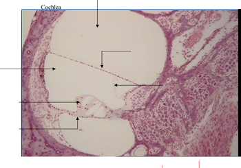
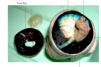
Label arrows.
First picture is Cochlea
SEcond is Cow eye

Transcribed Image Text:This histological image shows a section of respiratory epithelium, illustrating the ciliated pseudostratified columnar epithelium found in the trachea and upper respiratory tract. Each labeled component plays a specific role in maintaining respiratory health:
1. **Cilia**: These hair-like structures extend from the surface of ciliated cells. Their primary function is to move mucus and trapped particles out of the respiratory tract.
2. **Goblet Cell**: These are specialized epithelial cells responsible for secreting mucus. Mucus serves to trap dust, pathogens, and particles, preventing them from reaching deeper parts of the respiratory system.
3. **Basal Cell**: These cells act as progenitor cells, capable of dividing and differentiating into other types of epithelial cells, playing a critical role in the repair and regeneration of the epithelium.
4. **Basement Membrane**: This thin, fibrous layer underlies the epithelium, providing structural support and anchoring the epithelial cells to connective tissue.
5. **Lamina Propria**: The layer of connective tissue beneath the epithelium. It contains blood vessels, nerves, and immune cells essential for nutrient exchange and defense mechanisms.
The image provides a detailed visualization of healthy respiratory epithelium, essential for both understanding normal airway function and recognizing pathological changes in respiratory diseases.

Transcribed Image Text:The image showcases a dissected human eyeball with various parts labeled for educational purposes:
1. **Cornea and Sclera**: The outermost layer of the eye is visible. The cornea is transparent and prominently positioned, while the sclera is the white part surrounding it.
2. **Pupil and Iris**: Visible at the center of the eye. The pupil appears as a dark circle, surrounded by the iris, which is often colored.
3. **Aqueous Humor**: The fluid-filled space between the cornea and the lens area is indicated, although not directly visible in the image.
4. **Lens**: Positioned behind the pupil and iris, the lens appears transparent and is responsible for focusing light onto the retina.
5. **Retina and Choroid**: The back of the eye is highlighted, showing layers responsible for receiving light and housing blood vessels.
6. **Optic Nerve**: This is indicated at the back, which transmits visual information from the retina to the brain.
The dissection reveals the internal structure of the eye, providing insights into the components involved in the human visual system.
Expert Solution
This question has been solved!
Explore an expertly crafted, step-by-step solution for a thorough understanding of key concepts.
This is a popular solution
Trending nowThis is a popular solution!
Step by stepSolved in 2 steps with 2 images

Knowledge Booster
Learn more about
Need a deep-dive on the concept behind this application? Look no further. Learn more about this topic, biology and related others by exploring similar questions and additional content below.Similar questions
- Amniotes focus on nearby objects by _____. moving their lens forward in their eye further from the retina moving their lens backward in their eye closer to the retina contracting their ciliary muscle to release tension on the zonular fibers so that the lens becomes rounder contracting their ciliary muscle to increase tension on the zonular fibers so that their lens becomes flatter relaxing their ciliary muscle to put tension on the ciliary fibers so that their lens becomes flatterarrow_forwardName the part that is affected in astigmatism.arrow_forwardExplain how the diagram is too simple.arrow_forward
arrow_back_ios
arrow_forward_ios
Recommended textbooks for you
 Human Anatomy & Physiology (11th Edition)BiologyISBN:9780134580999Author:Elaine N. Marieb, Katja N. HoehnPublisher:PEARSON
 Biology 2eBiologyISBN:9781947172517Author:Matthew Douglas, Jung Choi, Mary Ann ClarkPublisher:OpenStax
 Anatomy & PhysiologyBiologyISBN:9781259398629Author:McKinley, Michael P., O'loughlin, Valerie Dean, Bidle, Theresa StouterPublisher:Mcgraw Hill Education,
 Molecular Biology of the Cell (Sixth Edition)BiologyISBN:9780815344322Author:Bruce Alberts, Alexander D. Johnson, Julian Lewis, David Morgan, Martin Raff, Keith Roberts, Peter WalterPublisher:W. W. Norton & Company
 Laboratory Manual For Human Anatomy & PhysiologyBiologyISBN:9781260159363Author:Martin, Terry R., Prentice-craver, CynthiaPublisher:McGraw-Hill Publishing Co.
 Inquiry Into Life (16th Edition)BiologyISBN:9781260231700Author:Sylvia S. Mader, Michael WindelspechtPublisher:McGraw Hill Education

Human Anatomy & Physiology (11th Edition)
Biology
ISBN:9780134580999
Author:Elaine N. Marieb, Katja N. Hoehn
Publisher:PEARSON

Biology 2e
Biology
ISBN:9781947172517
Author:Matthew Douglas, Jung Choi, Mary Ann Clark
Publisher:OpenStax

Anatomy & Physiology
Biology
ISBN:9781259398629
Author:McKinley, Michael P., O'loughlin, Valerie Dean, Bidle, Theresa Stouter
Publisher:Mcgraw Hill Education,

Molecular Biology of the Cell (Sixth Edition)
Biology
ISBN:9780815344322
Author:Bruce Alberts, Alexander D. Johnson, Julian Lewis, David Morgan, Martin Raff, Keith Roberts, Peter Walter
Publisher:W. W. Norton & Company

Laboratory Manual For Human Anatomy & Physiology
Biology
ISBN:9781260159363
Author:Martin, Terry R., Prentice-craver, Cynthia
Publisher:McGraw-Hill Publishing Co.

Inquiry Into Life (16th Edition)
Biology
ISBN:9781260231700
Author:Sylvia S. Mader, Michael Windelspecht
Publisher:McGraw Hill Education