Human Anatomy & Physiology (11th Edition)
11th Edition
ISBN: 9780134580999
Author: Elaine N. Marieb, Katja N. Hoehn
Publisher: PEARSON
expand_more
expand_more
format_list_bulleted
Question

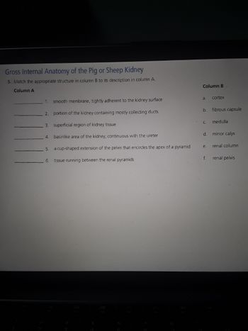
Transcribed Image Text:**Gross Internal Anatomy of the Pig or Sheep Kidney**
**Exercise 5:** Match the appropriate structure in column B to its description in column A.
<table>
<tr>
<th>Column A</th>
<th>Column B</th>
</tr>
<tr>
<td>1. smooth membrane, tightly adherent to the kidney surface</td>
<td>a. cortex</td>
</tr>
<tr>
<td>2. portion of the kidney containing mostly collecting ducts</td>
<td>b. fibrous capsule</td>
</tr>
<tr>
<td>3. superficial region of kidney tissue</td>
<td>c. medulla</td>
</tr>
<tr>
<td>4. basinlike area of the kidney, continuous with the ureter</td>
<td>d. minor calyx</td>
</tr>
<tr>
<td>5. a cup-shaped extension of the pelvis that encircles the apex of a pyramid</td>
<td>e. renal column</td>
</tr>
<tr>
<td>6. tissue running between the renal pyramids</td>
<td>f. renal pelvis</td>
</tr>
</table>
Explanation of columns:
**Column A:** This column lists descriptions of different parts of the internal structure of the pig or sheep kidney. These descriptions provide key characteristics of each structure to help in identifying them.
**Column B:** This column lists the names of parts and structures associated with the internal anatomy of the pig or sheep kidney.
**How to Use this Section:** Match the letter from Column B that best fits the description from Column A. This exercise helps in reinforcing the understanding of the anatomical locations and functions of various kidney structures.
**Anatomical Diagrams and Explanations:**
While the document itself does not contain graphs or diagrams, if we were to include such visuals, here's how they might be explained:
1. **Diagram of Kidney Cross-section:** A cross-sectional image of a kidney would show the cortex (outer layer), medulla (inner region), renal pelvis, minor calyces, and other structures.
2. **Detailed Labeling:** Each part of the kidney would be clearly labeled to correspond with the descriptions from the exercise, providing a visual
Expert Solution
This question has been solved!
Explore an expertly crafted, step-by-step solution for a thorough understanding of key concepts.
This is a popular solution
Trending nowThis is a popular solution!
Step by stepSolved in 2 steps

Knowledge Booster
Similar questions
- Place the following structurees of the internal kidney in the correct order in which urine passes through them. A. Major calyx B. Renal tubule C. Renal corpuscle D. Minor calyx E. Renal pelvisarrow_forward1. Begin in the blood vessel entering the kidney (renal artery), trace a molecule of water as it travels to nephron, gets filtered and processed into urine, and exits the body. 2a. Describe the involuntary micturition reflex (e.g. infant). b. What are the suspected mechanisms of voluntary control of urination?arrow_forwardRENAL STENOSIS 1. In brief, what causes this disease/condition? 2. What symptoms or complications occur due to this disease/condition? 3. Which of the 10 key kidney parts/regions are most directly involved in this disease/condition? 4. How would those parts/regions look different in this disease/condition?arrow_forward
- 1. Site of filtrate formation? 2. The structure that conveys the processed filtrate (urine) to the renal pelvis? 3. Blood supply that directly receives substances from the tubular cells? 4. The primary site of tubular reabsorption? 5. Two substances thta are routinely found in filtrate but not in the urine product? 6. Which has greater specific gravity: 1 ml of urine or 1 ml of distilled water?arrow_forwardIs this the correct answer of labeling the name the components of the male urinary system?arrow_forwardIndicate the type of nephridium shown in the figure above_____________________________ Name the labeled structures A (entire brown structure) & B and indicate the step in the excretory process occurring at each. Structure Name Process step A. B.arrow_forward
- 28. Which is correct? Last option is incorrectarrow_forwardThe correct pathway of urine from the renal pyramid to the outside of the body is: 1. Ureter 2. Lumen of bladder 3. Minor calyx 4. Major calyx 5. Renal papilla 6. Renal pelvis 7. Urethra 8. Renal sinus Question 34 options: 5, 3, 4, 6, 1, 2, 7 5, 4, 3, 6, 7, 2, 1 5, 3, 4, 6, 8, 1, 2, 7 5, 3, 4, 8, 1, 2, 7arrow_forwardThe following structures are within the kidney. Place them in order that fluid traveling through thee nephron (including blood before its filtered) hits that structure from where blood enters to where urine exits the kidney. 1. Juxtaglomerular apparatus 2. collecting duct 3. proximal tubule 4. podocytesarrow_forward
- 1. The location of the kidneys in the body is best described as being: In the pelvic cavity In the mediastinum retroperitoneal In the thoracic cavity 2. Place the structures of the renal tubule in the correct order from the start of filtration to completed urine in the collecting duct. descending (proximal) loop of Henle proximal convoluted tubule (PCT) distal convoluted tubule ascending (distal) loop of Henle Bowman's capsule 3. Place the structures of the blood flow in the nephron in the correct order. glomerulus afferent arteriole peritubular capillaries efferent arteriole cortical radiate veinsarrow_forwardThe following structures articulate with the kidneys at the hilum: D - major calyx B - renal veins A and B A - renal arteries A, B, and C C - ureter A, B, C, and Darrow_forwardA 75 year old man comes to the physician because of a six-week history of poor urinary stream and inability to void completely. Physical examination shows a markedly enlarged prostate. The diagnosis of benign prostatic hyperplasia is made. Between which of the following regions of the urethra is obstruction of urine flow most likely located in this patient? The bladder neck and the membranous urethra The membranous urethra and the proximal bulb of the penis The peritoneal membrane and the levator ani The proximal bulb of the penis and the perineal membrane The trigone and the internal urethral orificearrow_forward
arrow_back_ios
SEE MORE QUESTIONS
arrow_forward_ios
Recommended textbooks for you
 Human Anatomy & Physiology (11th Edition)Anatomy and PhysiologyISBN:9780134580999Author:Elaine N. Marieb, Katja N. HoehnPublisher:PEARSON
 Anatomy & PhysiologyAnatomy and PhysiologyISBN:9781259398629Author:McKinley, Michael P., O'loughlin, Valerie Dean, Bidle, Theresa StouterPublisher:Mcgraw Hill Education,
 Human AnatomyAnatomy and PhysiologyISBN:9780135168059Author:Marieb, Elaine Nicpon, Brady, Patricia, Mallatt, JonPublisher:Pearson Education, Inc.,
 Anatomy & Physiology: An Integrative ApproachAnatomy and PhysiologyISBN:9780078024283Author:Michael McKinley Dr., Valerie O'Loughlin, Theresa BidlePublisher:McGraw-Hill Education
 Human Anatomy & Physiology (Marieb, Human Anatomy...Anatomy and PhysiologyISBN:9780321927040Author:Elaine N. Marieb, Katja HoehnPublisher:PEARSON

Human Anatomy & Physiology (11th Edition)
Anatomy and Physiology
ISBN:9780134580999
Author:Elaine N. Marieb, Katja N. Hoehn
Publisher:PEARSON

Anatomy & Physiology
Anatomy and Physiology
ISBN:9781259398629
Author:McKinley, Michael P., O'loughlin, Valerie Dean, Bidle, Theresa Stouter
Publisher:Mcgraw Hill Education,

Human Anatomy
Anatomy and Physiology
ISBN:9780135168059
Author:Marieb, Elaine Nicpon, Brady, Patricia, Mallatt, Jon
Publisher:Pearson Education, Inc.,

Anatomy & Physiology: An Integrative Approach
Anatomy and Physiology
ISBN:9780078024283
Author:Michael McKinley Dr., Valerie O'Loughlin, Theresa Bidle
Publisher:McGraw-Hill Education

Human Anatomy & Physiology (Marieb, Human Anatomy...
Anatomy and Physiology
ISBN:9780321927040
Author:Elaine N. Marieb, Katja Hoehn
Publisher:PEARSON